Vật lý trị liệu
Vật lý trị liệu
Môn: Vật lý trị liệu (7720603) 10 tài liệu
Trường: Trường Đại học Kỹ thuật Y tế Hải Dương 13 tài liệu
Tác giả:


Preview text:
lOMoARcPSD|36041561
Theraputic Equipments Assist. Lec. Ra’aed Alobaidi Micro Wave Diathermy
The diathermies have been used in the treatment of a variety of musculoskeletal conditions,
including muscle strains, contusions, ligament sprains, tendinitis, tenosynovitis, bursitis, joint
contractures, myofascial trigger points, and osteoarthritis.
Technique of application of micro wave diathermy
1. Preparation of apparatus.
A. The selected emitter is connected to the machine by the appropriate cable and the power switched on.
B. There will be some delay before out put obtained. The physiotherapist tests the
apparatus by placing his or her hand in front of the emitter.
C. Increases the output until a sensation of warmth is experienced.
D. The controls are then returned to zero and the switch turned to the stand up position.
2. preparation of the patient
a) Evaluate the patient’s problem and determine the goals of treatment.
b) Determine that diathermy is not contraindicated.
c) Explain the procedure and the reason for applying diathermy to the patient and the
sensations the patient can expect to feel.
d) During application of thermal level diathermy, the patient should feel a comfortable
sensation of mild warmth with no increase in pain or discomfort.
e) The patient should be advised to move as little as possible during the treatment because
the strength of the field will change if the distance between the applicator and the treatment area changes.
f) Remove all metal jewelry and clothing from the area to be treated.
g) Position the patient comfortably on a chair or plinth with no metal components.
h) Sensitivity of the skin must be tested for the heat and cold.
3. Application of emitter.
The emitter is arranged so that its surface is parallel to the skin and at the appropriate Raaed Alobaidi
distance, due consideration being given to the surface marking of the structure to be
treated. Irregular surfaces and areas which perspire freely should, if possible, be avoided. 4. Irradiation
a. The patient is reminded of the sensation to be expected and of the need to report
accurately on that experienced.
b. The output is increased slowly until a sensation of warmth is experienced or the selected output is reached. 1
Downloaded by Nga T??ng (ngahuong55@gmail.com) lOMoARcPSD|36041561
Theraputic Equipments Assist. Lec. Ra’aed Alobaidi
c. The irradiation continues for an appropriate time.
d. The physiotherapist visiting the patient frequently to ensure that nothing untoward has occurred.
e. Slight erythema may be observed, but there should be no marked skin reaction.
f. When the treatment is complete, turn off the device, remove the applicator , and inspect
the treatment area. It is normal for the area to appear slightly red, and it may also feel warm to the touch. 5. Dosage
a) The dosage can be calculated from the power output of the machine, which may be up to 200 watts.
b) In all cases the sensation experienced by the patient must by primary guide when giving doses.
c) As a general rule, weaker doses should be used for acute than for chronic conditions.
d) The duration of irradiation ranges from 10 to 30 minutes.
e) It s advisable to commence cautiously and in all cases progressive increases in exposure
must depend on the patients reaction.
f) Treatment may be given daily or on alternate days. Raaed Alobaidi 2
Downloaded by Nga T??ng (ngahuong55@gmail.com) lOMoARcPSD|36041561
Therapeutic Equipment’s Assist. Lect. Ra’aed Alobaidi
Pulsed shortwave diathermy (PSWD)
Has also been used for its non-thermal effects in the treatment of soft-tissue injuries and wounds.
The mechanism of its effectiveness has been theorized to occur at the
cellular level, relating specifically to cell membrane potential.
Damaged cells undergo depolarization, resulting in cell dysfunction that
might include loss of cell division and proliferation and loss of regenerative capabilities.
Pulsed shortwave diathermy has been said to repolarize damaged cells, thus correcting cell dysfunction.
It has also been suggested that sodium tends to accumulate in the cell
because of a decrease in activity of the sodium pump during the
inflammatory process, thus creating a negatively charged environment.
When a magnetic field is induced, the sodium pump is reactivated, thus
allowing the cell to regain normal ionic balance.
pulsed electromagnetic energy (PEME) , pulsed electromagnetic field
(PEMF) , or pulsed electromagnetic energy treatment (PEMET) , is a
relatively new form of diathermy
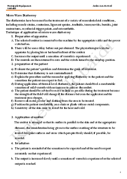
Application Techniques for Pulsed Shortwave Therapy 1. Parameters
Pulse duration is short, ranging from 20 to 400 sec
Pulse repetition rate, which ranges between 1 and 7000 Hz.
Peak pulse power the amplitude pulse/intensity 100-1000W.
Mean power power delivered from the machine (during pulse and average) Raaed Alobaidi 2. Preparation of the patient 1
Downloaded by Nga T??ng (ngahuong55@gmail.com) lOMoARcPSD|36041561
Therapeutic Equipment’s Assist. Lect. Ra’aed Alobaidi
Evaluate the patient’s problem and determine the goals of treatment.
Confirm that diathermy is the most appropriate intervention. Non-thermal
PSWD can be used where heat is contraindicated or potentially hazardous
and can be applied to insensate patients or to those who cannot tolerate the
sensations associated with other physical agents such as cryotherapy or electrical stimulation.
Determine that diathermy is not contraindicated.
Explain the procedure and the reason for applying diathermy to the patient
and the sensations the patient can expect to feel. The application of non-
thermal PSWD is not generally associated with any change in patient
sensation, although some patients report feeling slight tingling or mild warmth.
Remove all metal jewelry and clothing from the area to be treated.
Clean and dry the skin, and inspect it if necessary.
If applying non-thermal PSWD, it is not necessary to place towels between
the applicator and the body, but a disposable cloth or plastic covering can be
used over the applicator when treating conditions in which there is risk of
cross-contamination or infection.
3. Preparation the apparatus
Tune the device. Most modern diathermy devices tune automatically. To
tune a device that requires manual tuning, first turn it on and allow it to
warm up then turn up the intensity to a low level, and adjust the tuning dial
until a maximal reading on the power/intensity indicator is obtained.
Select the appropriate treatment parameters. When applying non thermal
PSWD, most clinicians select the intensity, pulse frequency, and total
treatment time based on the manufacturer’s recommendations, most non
thermal PSWD treatments are administered for 30 to 60 minutes once or
twice a day, 5 to 7 times a week. 4. Starting treatment Raaed Alobaidi
Provide the patient with a bell or other means to call for assistance during
treatment and if he or she experiences excessive heating or an increase in pain or discomfort.
After 5 minutes, check to be certain that the patient is not too hot or is not
experiencing any increase in symptoms.
When the treatment is complete, turn off the device, remove the applicator
and towels, and inspect the treatment area. It is normal for the area to appear
slightly red, and it may also feel warm to the touch.
Assess the outcome of the intervention. 2
Downloaded by Nga T??ng (ngahuong55@gmail.com) lOMoARcPSD|36041561
Therapeutic Equipment’s Assist. Lect. Ra’aed Alobaidi
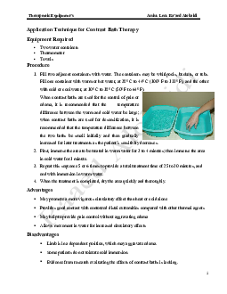
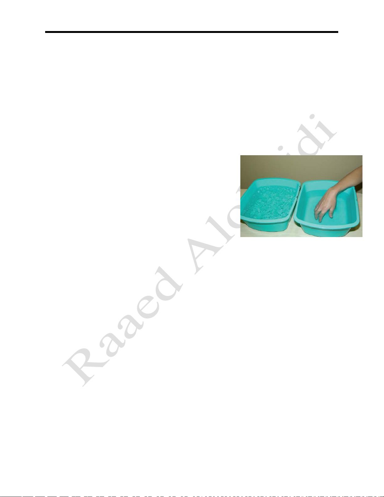
Application Technique for Contrast Bath Therapy Equipment Required
• Two water containers • Thermometer • Towels Procedure
1. Fill two adjacent containers with water. The containers may be whirlpools, buckets, or tubs.
Fill one container with warm or hot water, at 38° C to 44° C (100° F to 111° F), and the other
with cold or cool water, at 10° C to 18° C (50° F to 64° F).
When contrast baths are used for the control of pain or
edema, it is recommended that the temperature
difference between the warm and cold water be large;
when contrast baths are used for desensitization, it is
recommended that the temperature difference between
the two baths be small initially and then gradually
increased for later treatments as the patient’s sensitivity decreases.
2. First, immerse the area to be treated in warm water for 3 to 4 minutes; then immerse the area in cold water for 1 minute.
3. Repeat this sequence 5 or 6 times to provide a total treatment time of 25 to 30 minutes, and
end with immersion in warm water.
4. When the treatment is completed, dry the area quickly and thoroughly. Advantages
• May promote a more vigorous circulatory effect than heat or cold alone • Raaed Alobaidi
Provides good contact with contoured distal extremities compared with other thermal agents
• May help to provide pain control without aggravating edema
• Allows movement in water for increased circulatory effects Disadvantages
• Limb is in a dependent position, which may aggravate edema.
• Some patients do not tolerate cold immersion.
• Evidence from research evaluating the effects of contrast baths is lacking. 1
Downloaded by Nga T??ng (ngahuong55@gmail.com) lOMoARcPSD|36041561
Therapeutic Equipment’s Assist. Lect. Ra’aed Alobaidi
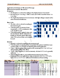
Application Technique of Ultrasound Therapy
Ultrasound Treatment Parameters Frequency
The frequency is selected according to the depth of tissue to be treated.
For tissue up to 5 cm deep, 1 MHz is used, and 3 MHz is used for tissue 1 to 2 cm deep.
The depth of penetration is lower in tissues with high collagen content and in areas of increased reflection. Duty Cycle
The duty cycle is selected according to the treatment goal.
When the goal is to increase tissue
temperature, a 100% (continuous) duty cycle should be used.
When ultrasound is applied where only
the non-thermal effects without tissue
heating are desired, pulsed ultrasound
with a 20% or lower duty cycle should be used. Intensity
o Intensity is selected according to the treatment goal.
o When the goal is to increase tissue temperature, the patient should feel some
warmth within 2 to 3 minutes of initiating ultrasound application.
o When 1 MHz frequency ultrasound is used, an intensity of 1.5 to 2.0 W/cm2
will generally produce this effect.
o When 3 MHz frequency is used, an intensity of about 0.5 W/cm2 is generally sufficient. o Raaed Alobidi
For non-thermal effects, successful treatment outcomes have been
documented for most applications using an intensity of (0.5 to 1.0 W/cm2),
with as low as 0.15 W/cm2 sufficient for facilitation of bone healing. 1
Downloaded by Nga T??ng (ngahuong55@gmail.com) lOMoARcPSD|36041561
Therapeutic Equipment’s Assist. Lect. Ra’aed Alobaidi Duration of Treatment
The length of the treatment is dependent on several factors:
1. The size of the area to be treated; 2. The intensity in W/cm2; 3. The frequency;
4. The desired temperature increase.
Typically recommended treatment times have ranged between 5 and 10 minutes in length.
When the goal of treatment is to increase tissue temperature, the treatment duration
should be adjusted according to the frequency and intensity of the ultrasound.
Ultrasound rate of heating per minutes Exposure Techniques Direct Contact
Involves actual contact between the applicator and the
skin, a layer of gel should be applied to the treatment area
in sufficient amounts to maintain good contact and
lubrication between the transducer and the skin. Immersion (water bath)
The immersion technique is recommended if the area to
be treated is smaller than the diameter of the available
transducer or if the treatment area is irregular with bony Raaed Alobidi
prominences. A plastic, ceramic, or rubber basin should be
used, A basin filled with de-gassed water is used if
possible .Ordinary tap water presents the problem that gas
bubbles dissociate out from the water, accumulate on the
patients skin and the treatment head, and reflect the
ultrasonic beam. If tap water has to be used then the gas
bubbles must be wiped from these surfaces frequently
.The technique of application is that the treatment head is
held 0.5 - 1 cm from the skin and moved in small concentric circles. 2
Downloaded by Nga T??ng (ngahuong55@gmail.com) lOMoARcPSD|36041561
Therapeutic Equipment’s Assist. Lect. Ra’aed Alobaidi Bladder Technique
A bladder technique can be used in which a balloon,
surgical glove can be filled with water, and the
ultrasound energy is transmitted from the transducer
to the treatment surface through this bladder.
Both sides of the balloon should be coated with gel
to assure better contact. Treatments using a bladder
filled with either gel or silicone or degassed water
have also been used at higher ultrasound intensities. Commercial gel packs
Have gained popularity and several studies have
demonstrated their efficacy as a coupling medium.
When ultrasound is applied over bony prominences,
a gel pad should be covered with ultrasound gel on
both sides to ensure optimal heating.
Techniques of application Equipment Required • Ultrasound unit
• Gel, water, or other transmission medium • Antimicrobial agent • Towel
Evaluate the patient’s clinical findings and set the goals of treatment.
Confirm that ultrasound is not contraindicated for the patient or the condition.
Preparation of the patient and apparatus before use:-
1. Put the patient in comfortable position;
2. Remove the clothes from the area to be treated; 3. Raaed Alobidi
Select the optimal treatment parameters, including Ultrasound frequency, intensity, duty cycle, and duration;
the appropriate size of the treatment area; and the appropriate number and frequency of treatments. 3
Downloaded by Nga T??ng (ngahuong55@gmail.com) lOMoARcPSD|36041561
Therapeutic Equipment’s Assist. Lect. Ra’aed Alobaidi
Inspect the skin of the treated area:- 1. skin must be clean and dry;
2. sensitivity of the skin must be tested if there is hypersensitivity, by using very small dose;
3. Before treatment of any area with a risk of cross-infection, swab the sound
head with 0.5% alcoholic chlorhexidine, or use the antimicrobial approved for this use in the facility.
Explain the work of the apparatus to the patient:-
1) Tell the patient about the period of treatment.
2) Not necessary the patients feel any think during treatment.
3) If the patient feels pain in the treated area he must tell the physiotherapist.
If the patient complain of pain during the session you must:- 1) Increase the amount of gel; 2) Decreases the dosage;
3) Stop the session if the pain continues.
Apply an ultrasound transmission medium to the area to be treated.
1) Apply enough medium to eliminate any air between the sound head and the treatment area.
2) Select a medium that transmits ultrasound well, does not stain, is not
allergenic, is not rapidly absorbed by the skin, and is inexpensive.
Gels or lotions meeting these criteria have been specifically
formulated for use with ultrasound.
3) Or, for the application of ultrasound under water, place the area to be
treated in a container of water
Place the sound head on the treatment area and turn on the ultrasound Raaed Alobidi machine.
Move the sound head within the treatment area.
The sound head is moved at approximately 4 cm/second quickly enough to
maintain motion and slowly enough to maintain contact with the skin.
When the intervention is completed, remove the conduction medium from
the sound head and the patient, and reassess for any changes in status. 4
Downloaded by Nga T??ng (ngahuong55@gmail.com)