Smooth Muscle | Tài liệu Tiếng Anh
Smooth Muscle | Tài liệu Tiếng Anh . Tài liệu giúp bạn tham khảo, ôn tập và đạt kết quả cao. Mời đọc đón xem!
Môn: Tiếng Anh chuyên ngành 332 tài liệu
Trường: Tài liệu Tiếng Anh chuyên ngành, Tiếng Anh cho người đi làm 432 tài liệu
Tác giả:
Preview text:
C. Smooth Muscle I. Characteristics
- Located In the walls of hollow organs, blood vessels, eye, glands, uterus, skin .
- Control ed involuntarily by endocrine and autonomic nervous systems .
- Constriction of organs and vessels are slow and uniform (e.g., Peristalsis of intestinal tract;
Vasoconstriction of b.v. and other structures).
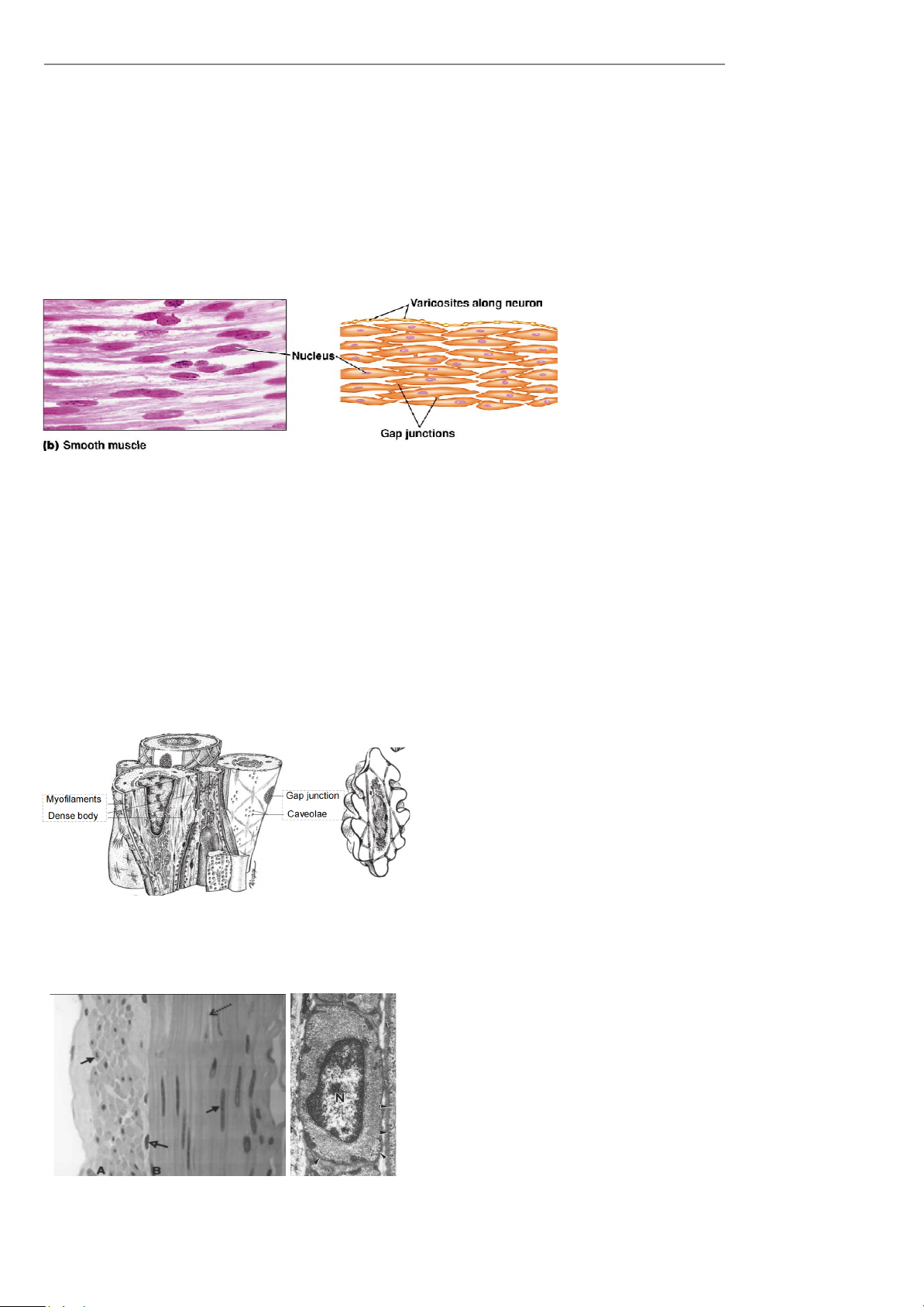
- Spindle-shaped cel s – 1 central nucleus. They range from 5 to 20 µm in diameter & from 20 µm to 1 mm/more in length.
- Activation is involuntary and it is fatigue resistant .
- Grouped into sheets: often running perpendicular to each other. II.
Functions and structure 1. Functions
- Propel urine, mix food in digestive tract, regulate blood flow.
- T o alter the activity of various body parts to meet the needs of the body at that time. 2. Structure
- Fibers are smal er than those in skeletal muscle:
+ Thin myofilaments: actin > tropomyosin (lack of troponin).
+ Thick myofilaments: myosin-II (spare).
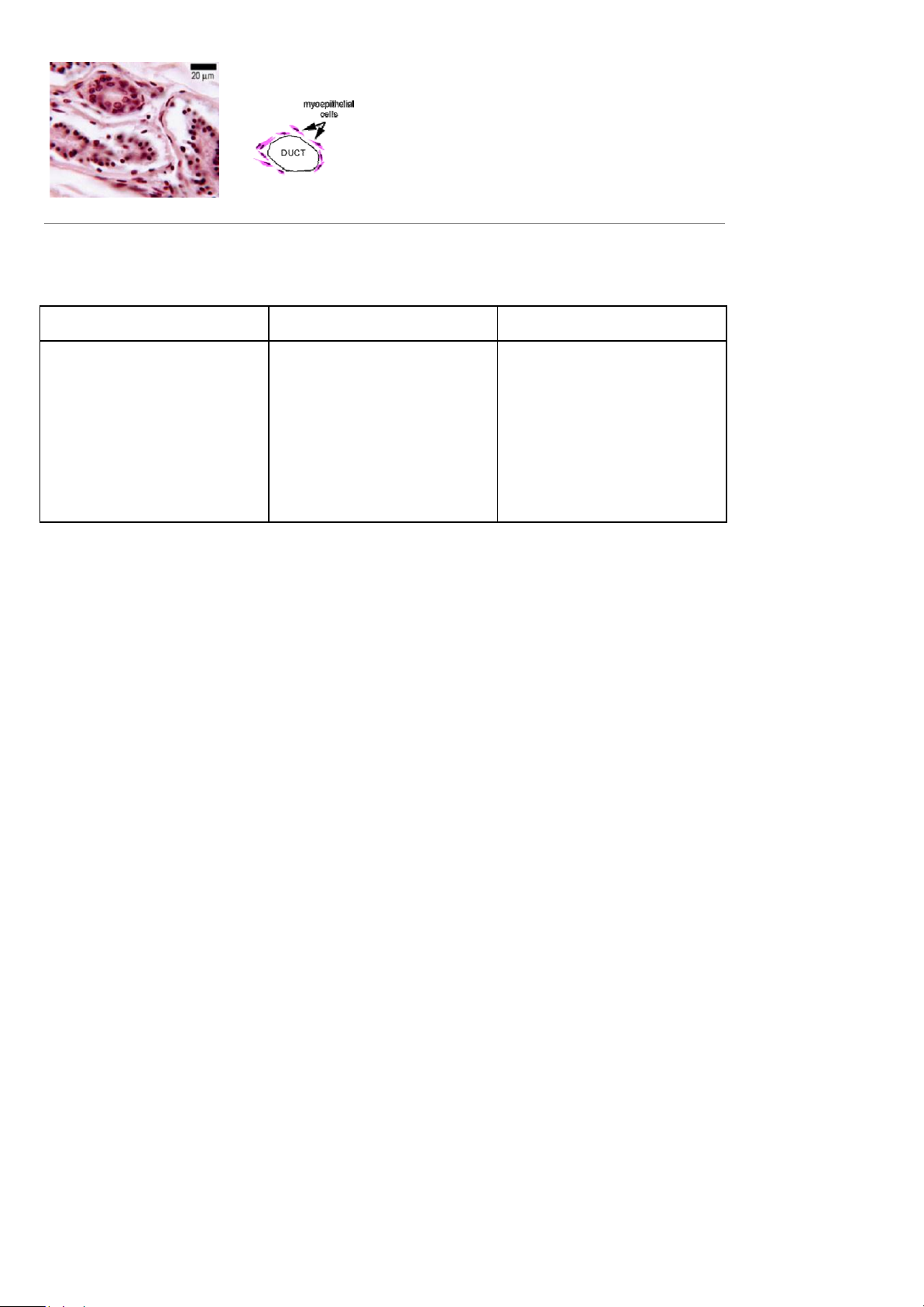
+ Intermediate filaments (desmin and vimentin) further link the dense bodies into a meshwork array.
- No sarcomeres . Not arranged as symmetrically as in skeletal muscle, thus NO striations .
- Dense bodies (instead of Z disks): serve as anchor sites for the myofilaments.
- Caveolae (indentations in sarcolemma) may act like T tubules: play a role in calcium transport.
- The smooth muscle cel has a central y located nucleus surrounded by cytoplasm containing
myofilaments in various orientations..
- When the myofilaments contract, the cel shortens
- Does not always require a nervous signal: can be stimulated by stretching or hormones .
Smooth muscle. A . Cross section. B. Longitudinal section.
The central myocyte nuclei (solid arrows) are absent in
several cross sections due to sectional geometry. The tip of a
spindle-shaped cel is visible at the dotted arrow. Fibroblast
nuclei (open arrow) are dark and smal er than smooth muscle
nuclei. Hematoxylin and eosin (×490)
Electron micrograph of a cross sectioned smooth muscle
cell. The nucleus (N) is centrally located, and the
cytoplasm contains numerous myofilaments.
Electron-dense bodies (*) serve as attachment sites for
the myofilaments. Numerous caveolae (arrowheads) are
present along the plasma membrane of an adjacent cell.
The basal laminae (L) are visible between the two cells
and appear fused at points (×23,900). (Courtesy of W. S. Tyler.)
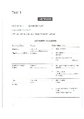
- Myo-epithelial cells: Single
smooth muscle cel s are often
found surrounding ducts, or blood
vessels, lying within the basement
membrane. They’re parts of the epithelial layer
D. Regeneration of Muscle and properties of Muscular Tissue I. Regeneration: Skeletal muscle Cardiac muscle Smooth muscle
- Skeletal muscle fibers - Cardiac muscle fibers - Smooth muscle fibers
cannot divide after 1st year .
cannot divide or regenerate ; regeneration is possible
- Growth is enlargement of al healing is done by fibrosis - Cel s can grow in size existing cel s. (scar formation) . (hypertrophy)
- Repair : satel ite cel s & bone - Some cel s (uterus) can marrow produce some new divide (hyperplasia)
cel s; if not enough numbers, - New fibers can form from fibrosis occurs most often stem cel s in BV wal s II.
Properties of Muscular Tissue:
1. Excitability - Ability to respond to stimuli.
2. Contractility - Ability to contract/shorten forceful y when stimulated.
3. Extensibility - Ability to stretch without being damaged.
4. Elasticity - Ability to return to an original length. III.
Functions of muscular system
- Producing Body Movements (Walking and running)
- Stabilizing Body Positions (Posture)
- Moving Substances Within the Body (blood, lymph, urine, air, food and fluids, sperm)
- Generating heat: contracting muscle produces heat; shivering increases heat production
Tài liệu liên quan:
-
Cambridge IELTS 13 Test-1 Overview and Practice Review | Tài liệu Tiếng Anh
5 3 -
IELTS Test 2 Review - Cambridge 13 Preparation Guide | Tài liệu Tiếng Anh
7 4 -
Litfocus Litreading Survey: Student Reading Interests & Habits Questionnaire | Tài liệu Tiếng Anh
6 3 -

Sách Pre-intermediate Market leader - Business English Course Book
22 11 -

Exam booster for B1 preliminary and B1 preliminary for schools with answer key
8 4