Báo cáo lâm sàng: Ngộ độc thuốc diệt chuột nhóm chống đông Bromadiolone
Báo cáo lâm sàng: Ngộ độc thuốc diệt chuột nhóm chống đông Bromadiolone. Tài liệu được sưu tầm giúp bạn tham khảo, ôn tập và đạt kết quả cao. Mời bạn đọc đón xem.
Môn: Tài liệu Tổng hợp 3.6 K tài liệu
Trường: Tài liệu khác 3.9 K tài liệu
Tác giả:


Preview text:
VETERINARSKI ARHIV 75 (3), 273-282, 2005
Intoxication with anticoagulant rodenticide bromadiolone in a dog - a case report
Rumen Binev*, Petko Petkov, and Anton Rusenov
Faculty of Veterinary Medicine, Trakia University, Stara Zagora, Bulgaria
BINEV, R., P. PETKOV, A. RUSENOV: Intoxication with anticoagulant rodenticide
bromadiolone in a dog - a case report. Vet. arhiv 75, 273-282, 2005. ABSTRACT
A spontaneous intoxication caused by the anticoagulant rodenticide bromadiolone (Lanirat 0.005) in a
Caucasian mountain shepherd dog at the age of 18 months is described. During its hospitalization at the Clinic
of Internal Diseases, Faculty of Veterinary Medicine in Stara Zagora, clinical, laboratory, microbiological,
radiological and ultrasonographic studies were performed. It was found that rodenticide intoxication was
manifested by changes in the clinical status (arexia, polydipsia, hyperthermia, polypnea, dyspnea, tachycardia,
pale conjunctives, liquidothorax and ascites) and in studied laboratory parameters (oligochromaemia, erythropenia,
leukocytosis with neutrophilia and regenerative shift, hyperglycaemia, increased ALAT activities and prolonged PT, APTT and PIVKA).
Key words: intoxication, anticoagulant, rodenticide, bromadiolone, dog Introduction
Pesticides are used all over the world in different branches of agriculture, industry,
health care, etc. Their wide application and inadequate information about the means of their
use and their toxic effects pose a real hazard for the health of both animals and humans (MARONI et al., 2000).
From the aspect of ecological agriculture the control of harmful rodents (mice, rats,
field mice, etc) requires precise and adequate use of chemical means of restriction of their
population. To date, the use of baits containing active substances with various composition
and anticoagulant effect is the most frequent of such means (SENGALEVICH et al., 1998). * Contact address:
Dr. Rumen Binev, Department of Internal Diseases and Clinical Toxicology, Faculty of Veterinary Medicine, Trakia University,
6000 Stara Zagora, Bulgaria, Phone: +359 42 2801 2658; Fax: 359 4274125; E-mail: binev@abv.bg ISSN 0372-5480 Printed in Croatia 273
R. Binev et al.: Intoxication with anticoagulant rodenticide bromadiolone in a dog - a case report
Anticoagulant rodenticides are probably the most commonly used of such means
around the world. It has been estimated that approximately 95% of all rodenticides used
are anticoagulant baits (MURPHY, 2002).
Anticoagulant rodenticides are classified into two principal groups: derivatives of
coumarin and indanedione. The preparations from the first group are more extensively used
and provoke severe injury to vascular permeability, resulting in massive haemorrhages and
the rapid death of rodents (SAMAMA et al., 2002; MURPHY, 2002; RADI and THOMPSON, 2004).
The mechanism of intoxication with coumarin anticoagulants is via a specific inhibition
of blood coagulation. Vitamin K is needed for the functional synthesis of coagulation factors
II, VII, IX and X. The most common vitamin K-responsive coagulopathy is anticoagulant
rodenticide intoxication (ROZANSKI et al., 1999; SAMAMA et al., 2002; MISCHKE and
NOLTE, 1999; MOUNT et al., 2003). Blood vessels lose their elasticity, and subsequently
ruptures of large blood vessels occur, clinically manifested by massive haemorrhages and
haematomes (PETRUS and HENIK, 1999; KOHN et al., 2003; LUTZE et al., 2003; MUNDEY and
THOMPSON, 2003; RADI and THOMPSON, 2004). PETTERINO and PAOLO (2001) showed
that anticoagulant rodenticides impair the cellular recirculation of vitamin K, causing secondary coagulopathies.
Up until now cases of intoxication with anticoagulant rodenticides and their effects
have been described in cats (KOHN et al., 2003), polecats (SHORE et al., 2003), in wild
animals (deer, coons, marten, eagle, owlet, hawk, etc.) with various anticoagulants such
as bromadifacoum, chlorophacinone, bromadiolone and coumatetralyl (STONE et al., 1999; STONE et al., 2003).
A spontaneous intoxication with an unknown anticoagulant rodenticide in a dog has
been described by BLOCKER and ROBERTS (1999); ROZANSKI et al. (1999); PETRUS and
HENIK (1999); PETTERINO and PAOLO (2001); MURPHY (2002); MOUNT et al. (2003);
MUNDAY and THOMPSON (2003); LUTZE et al. (2003); PETTERINO et al. (2004).
One of the most commonly used rodenticides is bromadiolone, used in the composition
of the commercial preparation Lanirat 0.005. Bromadiolone is a second-generation
coumarin anticoagulant. It is used at a dose of 8-10% added to a bait consisting of 100 kg
ground grain, 1.5 l sunflower oil, 4 l water and 2 l Lanirat 0.005. Thus, it is used to attract
harmful rodents (SENGALEVICH et al., 1998).
In the available literature there are no complete clinical and laboratory data for
spontaneous intoxication with bromadiolone in dogs, which motivated our complex
clinical, laboratory, microbiological, radiological and ultrasonographic studies in order to
facilitate the diagnostics and the treatment of bromadiolone intoxications. 274
Vet. arhiv 75 (3), 273-282, 2005
R. Binev et al.: Intoxication with anticoagulant rodenticide bromadiolone in a dog - a case report Materials and methods
On March 11, 2002 an 18-month-old male Caucasian shepherd dog from Shoumen
named Spike and weighing 60 kg was referred to the Clinic of Internal Diseases, Faculty
of Veterinary Medicine, Trakia University, Stara Zagora. Anamnesis revealed that a week
earlier the owner noticed a symmetrical abdominal enlargement, decreased appetite, frequent
urination and increased thirst. Three to four weeks previously a deratization by placement
of baits containing Lanirat 0.005 in farm premises, where the dog was used as a guard, had
been performed. For some time the dog was occasionally observed eating dead and dying
rodents. The animal’s diet normally consisted of granulated food and plenty of meat and fats.
Immunoprophylactic and antihelminthic drugs were regularly administered. During the
period prior to the referral, body temperature increased up to 40 oC. The dog was given
antibiotics: i.m. Shotapen and 2 000 000 UI Penicillin intraperitoneally, but with no effect.
During the hospitalization clinical status was followed twice daily by checking body
temperature (BT), heart rate (HR) and respiratory rate (RR), colour of mucosae, appetite,
thirst, general condition, locomotor activity, sensory perception, urination, defecation, etc.
applying routine clinical diagnostic methods.
At referral and at post-hospitalization days 4, 8, 12 and 16, blood from v. cephalica
antebrachii was sampled for determination of haemoglobin concentration (HGB), red blood
cell (RBC) counts, white blood cell (WBC) counts and haematocrit (HCT) (automated
haemoanalyzer Serono-System 150+, U.S.A.), and differential WBC counts by the method
of Pappenheim, erythrocyte sedimentation rate (ESR) by the method of Panchenko,
Thrombin time (TT), activated partial thromboplastine time (APTT), prothrombin time
(PT), protein induced by Vitamin K antagonism or absence (PIVKA) and activated clotting
time (ACT) using diagnostic kits (BIOLABO, France), and a coagulometer (Amelung,
Germany). The activities of transaminases ASAT and ALAT, uric acid, total bilirubin,
urea, creatinine and blood sugar were assessed using an automated analyser (Reflotron
Manual, Germany) using a Roche (Germany) diagnostic kit.
The course of the disease and its treatment were followed by thoracic and abdominal
radiography (TUR D-800-1, Germany) and ultrasonography probe (Aloka-SSD-500, UST-5871-5).
Several punctions of the thorax (in the left intercostal spaces VII-VIII and the right
intercostal spaces IV-V) and the abdomen were performed at hospitalization and post-
hospitalization, on days 3, 6, 9 and 12. Physical analysis of punctate included its colour,
transparency, density (using an aerometer), with a chemical analysis of the amount of
protein (Esbach’s albuminometer). The punctate was microscopically and microbiologically
studied on McConkey agar (blood agar with 5% sheep red blood cells). The Rivolta test
was applied for differentiating between transudate and exudate.
Vet. arhiv 75 (3), 273-282, 2005 275
R. Binev et al.: Intoxication with anticoagulant rodenticide bromadiolone in a dog - a case report Results
The physical examination revealed pale conjunctives, decreased locomotor activity
and sensory perception, rapid exhaustion, bilateral symmetrical abdominal enlargement
with drooping of the ventral wall and spinal lordosis. The patient was constantly sitting
up in order to ease respiration. Body temperature was 39.6 oC, heart rate 115 min-1 and respiratory rate – 88 min-1.
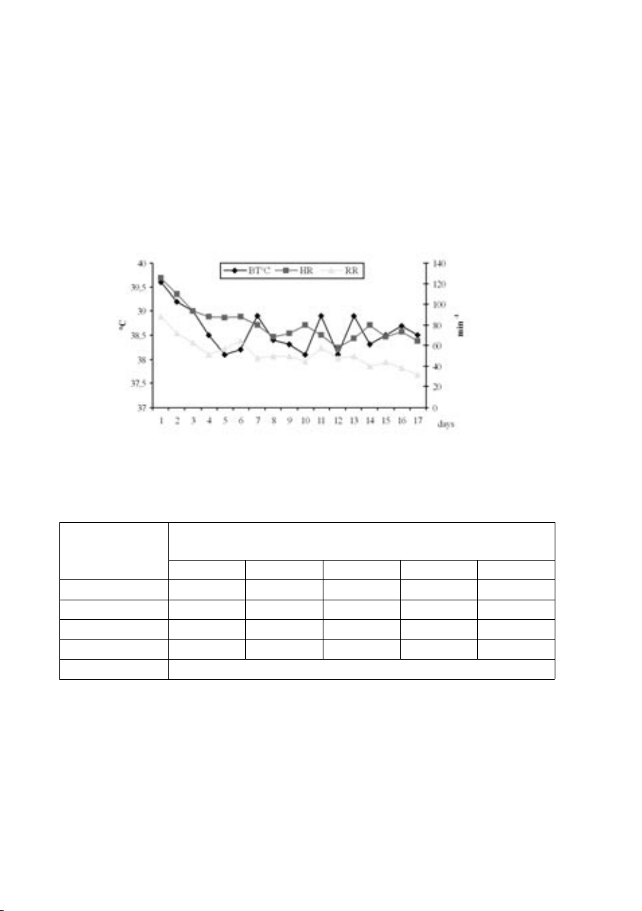
Fig. 1. Changes in body temperature (BT), heart rate (HR) and respiratory rate (RR) of a dog fol-
lowing intoxication with the anticoagulant rodenticide bromadiolone.
Table. 1. Results from the analysis of thoracic punctuates obtained from a dog following intoxica-
tion with anticoagulant rodenticide bromadiolone. Parameters Days after the hospitalization 1 3 6 9 12 Amount, mL 1700 500 650 400 250 Density 1,005 1,008 1,010 1,012 1,015 Protein g% 2 2.5 2.5 3 3 Rivolta’s test - - ± ± ± Sediment
Plenty of erythrocytes. Polymorphonuclear leucocytes and endothel cells
During hospitalization (Fig. 1) an elevated BT was present during the first two days,
but afterwards, until the end of the follow-up, it was within physiological range (37.5- 39.0 °C).
A tachycardia was observed from the first (115 min-1) until the 7th (82 min-1) day of
hospitalization. From day 8 until the end of follow-up, RR was normalized (60-80 min-1). 276
Vet. arhiv 75 (3), 273-282, 2005
R. Binev et al.: Intoxication with anticoagulant rodenticide bromadiolone in a dog - a case report
The pulse was strong, full, hard and rhythmic. After percussion a decrease in absolute
cardiac dullness was observed, while auscultation revealed stunt cardiac tones.
Breathing at referral was with increased frequency (88 min-1) difficult (with open
mouth) from a costo-abdominal type, with the active participation of abdominal muscles in
both respiratory phases. Percussion showed a dullness in the ventral pulmonary areas with
a horizontal upper limit that moved with lifting of the front part of the body. Auscultation
evidenced a bronchial respiration with moist fin bubbling rates over the limit of horizontal
dullness. Underneath, respiratory sounds were poorly audible. The RR was enhanced
(polypnea) for the whole period of observation (Fig. 1). The tendency towards lower RR
observed during the second half of the period did not result in normalization and the values
were still above physiological limits (15-25 min-1).
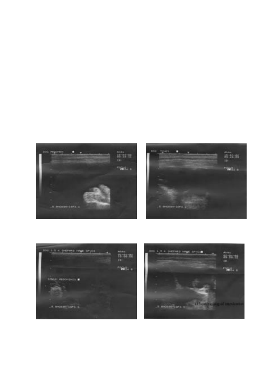
Fig. 2. Abdominal (left) and thoracic (right) ultrasonographies of a dog following intoxication
with anticoagulant rodenticide bromadiolone by day 1
Fig. 3. Abdominal (left) and thoracic (right) ultrasonographies of a dog following intoxication
with anticoagulant rodenticide bromadiolone by day 12
Vet. arhiv 75 (3), 273-282, 2005 277
R. Binev et al.: Intoxication with anticoagulant rodenticide bromadiolone in a dog - a case report
Table. 2. Clinical laboratory blood examinations data of a dog following intoxication with antico-
agulant rodenticide bromadiolone. Parameters Days after the hospitalization 1 4 8 12 16 Haematological parameters HGB g/L 82 88 98 115 122 RBC T/L 5.19 5.24 5.54 7.39 8.56 HCT % 33.3 35.7 43.4 55.3 61.1 WBC G/L 19.1 15.2 16.4 13.8 12.1 Differential wbc counts % Eo 0 0 1 1 0 Mm 3 5 3 4 2 St 18 25 14 13 22 Sg 70 64 65 77 62 Lym 8 6 16 5 14 Mo 1 0 1 0 0 Biochemical parameters Blood sugar mmol/L 6.48 6.23 6.87 5.54 4.82 Total bilirubin µmol/L 28.6 22.8 23.5 18.1 13.2 ASAT U/L 34.1 32.2 20.8 15.0 12.8 ALAT U/L 65.2 54.8 44.2 35.7 40.1 Urea mmol/L 18.8 12.4 9.6 12.8 10.2 Uric acid µmol/L 78,8 64,2 96,5 118,4 128,1 Creatinine µmol/L 148 124 109 111 94 PT s 122 84 36 18 14 APTT s 86 68 24 22 10 PIVKA s 148 96 45 32 15 TT s 18 10 14 15 10 ACT s 82 64 58 75 45
Lateral thoracic and abdominal radiographs were performed at referral, which revealed
shading with a horizontal upper limit. 278
Vet. arhiv 75 (3), 273-282, 2005
R. Binev et al.: Intoxication with anticoagulant rodenticide bromadiolone in a dog - a case report
Ultrasonography was also performed in order to assess the amount and position of liquid
in the thoracic and abdominal cavities (Fig. 2). Interpretation suggested vast anechogenic
spaces (filled with liquid), contrasting with the more echogenic zones of the liver, spleen,
intestines and the soft abdominal wall. A similar echogenic picture was also observed in the pleural cavity.
At day 12, a second ultrasonography was performed (Fig. 3) which evidenced a marked
decrease inf thoracic and abdominal liquids.
The optimal site of thoracic punction was found using general and special methods and
was located in the left VIIIth intercostal space in the zone of maximum dullness. During
hospitalization, 5 thoracic punctions were performed: in left intercostal spaces VII-VIII
and right intercostal spaces IV-V. The punctate had an intensively red colour and was
odourless. The results of analysed punctates are presented in Table 1, showing that they
were haemorrhagic transudates. The microbiologically studied was negative. Similar results
showed the data of the abdominal punctates.
Laboratory parameters, assessed during the hospitalization period (Table 2), showed
decreased HGB – 82 g/l, RBC – 5.19 T/l and HCT – 33.3% and increased WBC counts:
19.1 G/l. By the end of the observation those parameters returned to their physiological
values. The differential WBC counts suggested neutrophilia with a regenerative shift to Mm (3-5%).
Higher blood levels were measured for blood sugar, bilirubin and ALAT. They remained
elevated until the end of the hospitalization, although a tendency towards normalization
was observed. Significant deviations in the levels of ASAT, urea, creatinine, uric acid and
ESR were not observed during the period of study.
Prothrombin time (PT) was 122 s (reference range: 12-14 s), activated partial
thromboplastine time (APTT) was 88 s (reference range: 12-16 s) and protein induced
by vitamin K antagonism or absence (PIVKA) was 148 s (reference range: <25 s) at the
beginning of the period of observation. Following treatment it was restored to within the
reference range by day 12 of hospitalization. thrombin time (TT) and activated clotting
time (ACT) was in reference range. Discussion
Anticoagulant rodenticides impair the cellular recirculation of vitamin K causing
secondary coagulopathies, inhibits blood coagulation and affects blood vessels via an
analogous mechanism (BLOCKER and ROBERTS, 1999; PETRUS and HENIK, 1999; MUNDEY
and THOMPSON, 2003; KOHN et al., 2003). This explains haemocirculatory disorders
manifested by accumulation of haemorrhagic transudate in thoracic (liquidothorax) and
abdominal cavity (ascites) (RADI and THOMPSON, 2004), observed by us. It could be
Vet. arhiv 75 (3), 273-282, 2005 279
R. Binev et al.: Intoxication with anticoagulant rodenticide bromadiolone in a dog - a case report
presumed that the simultaneous decrease of RBC counts, haemoglobin (normochromic
anemia) and haematocrit values occurred as a consequence of blood loss. The observed
leukocytosis and hyperthermia are probably caused by immunodeficiency, which in turn
resulted in some kind of infection (KATARANOVSKI et al., 2003). Immunodeficiency
is a consequence of long-term anaemia and hypoxia, as well as liver injury (RADI and
THOMPSON, 2004). The large amount of thoracic transudate is most likely responsible for
the collapse of the compressed pulmonary parenchyma and for the signs of respiratory
insufficiency: polypnea, dyspnea, and costo-abdominal type of breathing. The tachycardia
is a compensatory mechanism for oligochromaemia, erythropenia and pulmonary
insufficiency. Hyperglycaemia, bilirubinaemia and increased ALAT activity are other
signs of the toxic effect of anticoagulant rodenticides on liver parenchyma. It could be
accepted that the same toxicodynamics are also valid for bromadiolone intoxication.
Treatment. Throughout the period of hospitalization (days 1-15), 300 mL 20% glucose,
5 mL 10% vitamin C, 5 mL 20% coffein natricum benzoicum intravenously and 3 mg/kg
body weight vitamin K subcutaneously were administered once daily. Medication therapy 1
was extended by intramuscular administration of 12 mL Lincomycin-Spectinomycin
5/10 (50 mg lincomycin hydrochloride and 100 mg spectinomycin dihydrochloride in
1 mL injectable solution) (Alfasan-Woerden, Holland) every 24 hours for 10 days; 20
mg Furosemide (in 2 mL injectable solution) every 12 hours for 3 days, followed by
oral application of 40 mg Furosemide tablets twice daily for 10 days, and 5 g urotropin
(hexamethylentetramin) once daily for 7 days.
The punctions of thoracic and abdominal cavities were followed by intrathoracal or
intraperitoneal administration of 10 mL vitamin AD E, (vitamin A 15000 UI, vitamin 3
D 20000 UI and vitamin E 0.01 g), 1 mL gentamicin (40 mg/mL), 5 mL 1% novokain 3
(1 g procaine hydrochloride in 100 mL, VetProm, Radomir, Bulgaria) and 1 mL
dexamethasone (2 mg dexamethasone disodium phosphate in 1 mL; Alfasan-Woerden, Holland).
After a 3-week therapy, the clinical and the laboratory status of the patient was within
reference ranges and the dog was discharged from the clinic as cured.
Anticoagulant rodenticide toxicosis is a potentially fatal condition, but it may be treated
successfully if diagnosis is made quickly and appropriate therapy is instituted. References
BLOCKER, T., B. ROBERTS (1999): Acute tracheal obstruction associated with anticoagulant
rodenticide intoxication in a dog. J. Small Anim. Pract. 40, 577-580.
KATARANOVSKI, M., M. VLASKI, D. KATARANOVSKI, N. TOSIC, S. MANDIC-RADIC,
V. TODOROVIC (2003): Immunotoxicity of epicutaneously applied anticoagulant rodenticide
warfarin: evaluation by contact hypersensitivity to DNCB in rats. Toxicology 188, 83-100. 280
Vet. arhiv 75 (3), 273-282, 2005
R. Binev et al.: Intoxication with anticoagulant rodenticide bromadiolone in a dog - a case report
KOHN, B., C. WEINGART, U. GIGER (2003): Haemorrhage in seven cats with suspected
anticoagulant rodenticide intoxication. J. Feline Med. Surg. 5, 295-304.
LUTZE, G., W. ROMHILD, J. ELWERT, J. LEPPET, K. KUTSCHMANN (2003): Phenprocoumon
(Marcumar, Falithrom) as an unusual reason for coumarin poisoning in a dog. Case report.
Dtsch. Tierärztl. Wochenschr.110, 31-33.
MARONI, M., C. COLOSIO, A. FERIOLI, A. FAIT (2000): Biological Monitoring of Pesticide
Exposure: a review. Introduction. Toxicology 143, 1-118.
MISCHKE, R., I. NOLTE (1999): Häemostasediagnostik beim Hund: 2. Prinzip, Technik und
Referenzberich verschidener Untersuchungsverfahren. Prak. Tierarzt 80, 836-855.
MOUNT, M. E., B. U. KIM, P. H. KASS (2003): Use of a test for proteins iduced by vitamin K
absence or antagonism in diagnosis of anticoagulant poisoning in dogs: cases (1987-1997). J.
Am. Vet. Med. Assoc. 222, 1070-1071.
MUNDEY, J. S., L. J. THOMPSON (2003): Brodifacoum toxicosis in two neonatal puppies. Vet. Pathol. 40, 216-219.
MURPHY, M. J. (2002): Rodenticides. Vet. Clin. Am. Small Anim. Pract. 32, 469-484.
PETRUS, D. J., R. A. HENIK (1999): Pericardial effusion and cardiac tamponade secondary to
brodifacoum toxicosis in a dog. J. Am. Vet. Med. Assoc. 215, 647-648.
PETTERINO, C., B. PAOLO (2001): Toxicolgy of various anticoagulant rodenticides in animals.
Vet. Hum. Toxicol. 43, 353-360.
PETTERINO, C., B. PAOLO, G. TRISTO (2004): Clinical and pathological features of anticoagulant
rodenticide intoxications in dogs. Vet. Hum. Toxicol. 46, 70-75.
RADI, Z. A., L. J. THOMPSON (2004): Renal subcapsular hematoma associated with brodifacoum
toxicosis in a dog. Vet. Hum. Toxicol. 46, 83-84.
ROZANSKI, E. A., K. J. DROBATZ, D. HUGHES, M. SCOTTI, U. GIGER (1999): Thrombotest
(PIVKA) Test Results in 25 Dogs with Acquired and Hereditary Coagulopathies. J. Vet. Emerg. Crit. Care 2, 73.
SAMAMA, M. M., G. T. GEROTZIAFAS, I. ELALAMY, M. H. HORELLOU, J. CONARD (2002):
Biochemistry and clinical pharmacology of new anticoagulant agents. Pathophysiol. Haemost. Thromb. 32, 218-224.
SENGALEVICH, G., T. TONEV, A. NIKOLOV (1998): Manual for Application of Chemicals for
Plant Protection, “Higher Agricultural Institute”, Plovdiv.
SHORE, R. F., J. D. BIRKS, C. L. WIENBURG, A. C. KITSCHENER (2003): Spatial and temporal
analysis of second-generation anticoagulant rodenticide residues in polecats (Mustela putorius)
from throughout their range in Britain, 1992-1999. Environ. Pollut. 122, 183-193.
STONE, W. B., J. C. OKONIEWSKI, J. R. STEDELIN (1999): Poisoning of wildlife with
anticoagulant rodenticides in New York. J. Wild. Dis. 35, 187-193.
STONE, W. B., J. C. OKONIEWSKI, J. R. STEDELIN (2003): Anticoagulant rodenticides and rapors:
recent findings from New York, 1998-2001. Bull. Environ. Contam. Toxicol. 70, 34-40. 281
R. Binev et al.: Intoxication with anticoagulant rodenticide bromadiolone in a dog - a case report Received: 17 January 2003 Accepted: 5 May 2005
BINEV, R., P. PETKOV, A. RUSENOV: Trovanje antikoagulantnim rodenticidom
bromadiolonom u psa - prikaz slučaja. Vet. arhiv 75, 273-282, 2005. SAŽETAK
Opisano je trovanje antikoagulantnim rodenticidom bromadiolonom (Lanirat 0,005) u kavkaskog planinskog
ovčara u dobi 18 mjeseci. U tijeku njegova liječenja provedene su kliničke, laboratorijske, mikrobiološke,
radiološke i ultrazvučne pretrage. Trovanje se očitovalo promjenama u kliničkoj slici (areksija, polidipsija,
hipertermija, polipneja, dispneja, tahikardija, bljedoća spojnica, likvidotoraks i ascites). Od laboratorijskih
pokazatelja ustanovljena je oligokromemija, eritropenija, leukocitoza s neutrofilijom i regenerativnim promjenama,
hiperglikemija, povećana aktivnost ALAT i produženo PT, APTT i PIVKA.
Ključne riječi: trovanje, antikoagulant, rodenticid, bromadiolon, pas 282
Vet. arhiv 75 (3), 273-282, 2005
Tài liệu liên quan:
-

Ung dung game hoa trong cac chien dich MKT
27 14 -
Bao cao Chi so TMDT Viet Nam 2025
30 15 -
Thông tư quy định về việc phân quyền, phân cấp và phân định thẩm quyền quản lý nhà nước về giáo dục cho chính quyền địa phương
31 16 -
Nghị quyết về phát huy các giá trị di sản văn hóa gắn với phát triên du lịch bền vững tỉnh Khánh Hòa đến năm 2025, định hướng đến năm 2030
33 17 -
Quyết định phê duyệt Chiến lược phát triển du lịch Việt Nam đến năm 2030
20 10