Lab 6 - Mitosis
Tài liệu báo cáo môn Bio for BME (BM090IU) tại Trường Đại học Quốc tế, Đại học Quốc gia Thành phố Hồ Chí Minh. Tài liệu gồm 11 trang giúp bạn ôn tập hiệu quả và đạt điểm cao! Mời bạn đọc đón xem!
Môn: Bio for BME (BM090IU) 7 tài liệu
Trường: Trường Đại học Quốc tế, Đại học Quốc gia Thành phố Hồ Chí Minh 2 K tài liệu
Tác giả:


Preview text:
lOMoARcPSD|364 906 32
VIETNAM NATIONAL UNIVERSITY – HO CHI MINH CITY
INTERNATIONAL UNIVERSITY
SCHOOL OF BIOMEDICAL ENGINEERING Biology for BME BM090IU REPORT LAB 6 Submitted by Date Submitted: 19/05/2023. Date Performed: 12/05/2023. Lab Section: Friday morning Course Instructor: Dr. Trinh Nhu Thuy
Assoc. Prof. Vong Binh Long, PhD lOMoARcPSD|364 906 32 Table of Contents
I. INTRODUCTION ....................................................................................................................... 1
1. Purpose .......................................................................................................................................... 1
2. Background information .......................................................................................................... 1
a. The Cell Cycle ......................................................................................................................... 1
b. Cell division - Mitosis ............................................................................................................. 2
II. EXPERIMENTAL .................................................................................................................... 3
1. Materials and equipment .............................................................................................................. 3
2. Experimental procedures ............................................................................................................. 3
III. RESULTS AND DISCUSSION ........................................................................................... 5
1. Results.............................................................................................................................................. 5
2. Discussion ........................................................................................................................................ 6
IV. CONCLUSION ......................................................................................................................... 6
International University BM090IU
School of Biomedical Engineering lOMoARcPSD|364 906 32 List of Figures
Figure 1. The cell cycle.. . .. . . . .. . . .. . . . .. . . .. . . .. . . .. . . . .. . . .. . . . .. . . . .. . . .. . . . .. . . 3
Figure 2. The comparision of division between animal and plant cell ... .. . .. . .. . .. . .... .5
Figure 3. Median longitudinal section of an onion root tip .. .. . .. . .. . .. . .. . .. . .. . . .. . .. . 6 List of Tables
Table 1. Required materials and equipment. . .. . . .. . . . .. . . . .. . . .. . . . .. . . .. . . . .. . .6
Table 2. Photographs of obsevation at 40x. .. ... .. . .. . .. . . .. . .. . .. . .. . .. . .. . .. . .. . . .. . .. . .. .7
International University BM090IU
School of Biomedical Engineering lOMoARcPSD|364 906 32 I. INTRODUCTION 1. Purpose
The goal of this experiment is to investigate the process of mitosis, or the division of the nucleus,
in eukaryotic cells. We will be able to examine and recognize the distinct stages of mitosis using a
microscope and prepared slides of onion root tips: prophase, metaphase, anaphase, and telophase.
This will aid our understanding of how cells multiply and retain genetic information. 2. Background information a. The Cell Cycle
The cell cycle, or cell-division cycle, is the series of events that take place in a cell that cause it to divide into two
daughter cells. These events include the duplication of its DNA (DNA replication) and some of
its organelles, and subsequently the partitioning of its cytoplasm, chromosomes, and other
components into two daughter cells in a process called cell division[1].
International University BM090IU
School of Biomedical Engineering 1 lOMoARcPSD|364 906 32
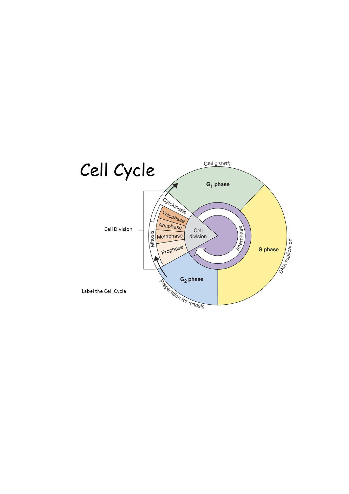
Figure 1. The Cell Cycle [2]
The cell cycle has four stages: G1, S, G2, and M. G1 and G2 are phases of cell growth and
preparation. S is the phase of DNA synthesis or replication. M is the phase of mitosis or cell division.
Interphase is the span between cell divisions that includes G1, S, and G2.[3]
G1 phase is one of the stages in which the interface of the life cycle of a cell is divided. Many
authors refer to this as the "growth phase", since during it the most significant growth of a cell
occurs. During the G1 phase, therefore, various intracellular metabolic changes occur that prepare the cell for division.[4]
S phase is the phase of the cell cycle where DNA synthesis or replication occurs. It happens during
interphase, before the cell divides by mitosis or meiosis. In S phase, the cell duplicates its entire
DNA and forms the centrosome, which helps to separate the DNA between the daughter cells. S
phase ensures that each daughter cell receives a complete copy of the genetic material of the parent cell.[5]
G2 phase, also known as Gap 2 phase or Growth 2 phase, is the third subphase of interphase in the
cell cycle. It follows the successful completion of S phase, during which the cell's DNA is
replicated. G2 phase is a period of rapid cell growth and protein synthesis during which the cell
prepares itself for mitosis. The cell replenishes its energy stores and synthesizes the proteins
necessary for chromosome manipulation. During G2 phase, the organism checks that any new cells
are not defective by verifying that the DNA has been replicated correctly and that there is enough
material present for two cells[6]. b. Cell division - Mitosis
In cell biology, mitosis is a part of the cell cycle in which replicated chromosomes are separated
into two new nuclei. Cell division by mitosis gives rise to genetically identical cells in which the
total number of chromosomes is maintained.[7][8] Therefore, mitosis is also known as equational division.
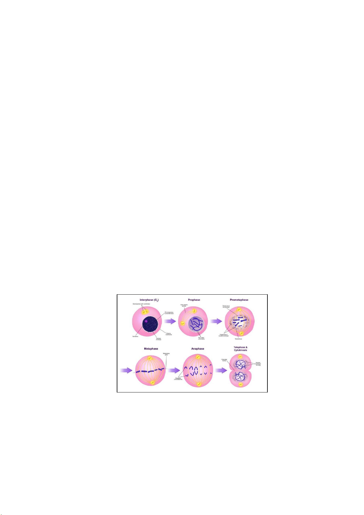
Figure 2. Phase of mitosis [9]
This phase is further divided into 4 phases, as follow:
Prophase (consists of prophase and prometaphase): the cell prepares to divide by tightly
condensing its chromosomes and initiating mitotic spindle formation. In prometaphase, the mitotic
spindle begins to capture and organize the chromosomes.
Metaphase: the spindle has captured all the chromosomes and lined them up at the middle of the cell, ready to divide.
International University BM090IU
School of Biomedical Engineering lOMoARcPSD|364 906 32
Anaphase: the sister chromatids separate from each other and are pulled towards opposite ends of the cell.
Telophase & Cytokinesis: The cell is nearly done dividing, and it starts to re-establish its normal
structures as cytokinesis (division of the cell contents) takes place. The division of the cytoplasm
to form two new cells overlaps with the final stages of mitosis. It may start in either anaphase or
telophase, depending on the cell, and finishes shortly after telophase.[10] II. EXPERIMENTAL 1. Materials and equipment
Table 1. List of requirements Materials
A stained slide of onion root tip cells Equipment Microscope 2. Experimental procedures
Experiment 1: Mitosis in Living Tissues – Onion Root Tips
Mitosis is easily studied using the root tips of actively growing plants. Roots of the common
garden onion (Allium cepa) provide good material for such a study (Figure 4). (Allium cepa has 16 chromosomes.)
International University BM090IU
School of Biomedical Engineering 3 lOMoARcPSD|364 906 32
Figure 3. Onion root[11]
Step 1: Obtain an onion root tip mitosis slide that was prepared.
Step 2: Place the slide onto the microscope stage and focus on the onion root tip at low power (10x,4x).
Step 3: Scan the tip to find areas of active cell division; this will be where we can observe mitosis.
Step 4: Once an area of cell division is found, switch to high power (40x) and observe the cells in the region.
Step 5: Identify each stage of mitosis in the cells you observed. Step
6: Record the observations and take pictures.
International University BM090IU
School of Biomedical Engineering lOMoARcPSD|364 906 32 III. RESULTS AND DISCUSSION 1. Results
Table 2. Photographs of observation at 40x Images from microscope Illustration Phase Interphases Prophases Metaphases
International University BM090IU
School of Biomedical Engineering 5 lOMoARcPSD|364 906 32 Anaphases Telophases 2. Discussion
Mitosis is the type of cell division that occurs in somatic cells, which are all the cells in the body
except for the reproductive cells (sperm and eggs). The purpose of mitosis is to create two
identical daughter cells that have the same number of chromosomes as the parent cell. This is
important for growth, tissue repair, and maintenance of the body. Mitosis is tightly regulated by
various proteins and checkpoints to ensure that each daughter cell receives the correct number
and type of chromosomes. Any errors or abnormalities during mitosis can result in genetic disorders or cancer. IV. CONCLUSION
The results of the experiment revealed the presence of all four stages of mitosis in the onion root
tip cells. The prophase stage was identified by the visible condensation of the chromatin into
chromosomes, and the disappearance of the nuclear membrane. In metaphase, the chromosomes
were seen aligning at the center of the cell. In anaphase, the chromosomes separated from each
other, and the spindle fibers pulled them towards opposite. In telophase, the chromosomes
decondensed and formed nuclei around them.
This lab report highlights the importance of the onion root tip as an excellent source of cells for
observing and studying the different stages of mitosis. The identification of all four stages of
mitosis, as well as the high mitotic index, affirmed our hypothesis that onion root tip cells are
continuously dividing. This experiment also teaches us the significance of mitosis in cell division
and growth, which is essential to maintain the growth and development of organisms.
International University BM090IU
School of Biomedical Engineering lOMoARcPSD|364 906 32 V. REFERENCES [1] Wikimedia Foundation. (2023, June 7). Cell cycle. Wikipedia.
https://en.wikipedia.org/wiki/Cell_cycle [2]
Karki, G. (2020, June 18). Phases of cell cycle. Online Biology Notes.
https://www.onlinebiologynotes.com/phases-of-cell-cycle/ [3]
Encyclopædia Britannica, inc. (2023, May 8). Cell cycle. Encyclopædia Britannica.
https://www.britannica.com/science/cell-cycle [4]
Morrison, R. (n.d.). G1 phase (cell cycle): Description and importance - science - 2023.
warbletoncouncil. https://warbletoncouncil.org/fase-g1-9142 [5]
Mayer, M. (2020, January 8). S phase: What happens during this subphase of the cell cycle?.
Sciencing. https://sciencing.com/s-phase-what-happens-during-this-subphase-of-the-cell- cycle13717820.html [6]
Wikimedia Foundation. (2022, December 19). G2 phase. Wikipedia.
https://en.wikipedia.org/wiki/G2_phase [7]
A. Laskey, R. (n.d.). Cell - Cell division and growth. Retrieved from
https://www.britannica.com/science/cell-biology/Cell-division-and-growth [8] Leacock, S. (2021). 4.1: Meiosis. Retrieved from
https://bio.libretexts.org/Courses/University_of_Arkansas_Little_Rock/Genetics_BIOL33
00_(Fa ll_2022)/Genetics_Textbook/04%3A_Inheritance/4.01%3A_Meiosis [9] Mitosis. (2022). Retrieved from
https://en.wikipedia.org/wiki/Mitosis#/media/File:Mitosis_Stages.svg [10]
Phases of Mitosis. (n.d.). Retrieved from https://www.khanacademy.org/science/ap-
biology/cell-communication-and-cellcycle/cell-cycle/a/phases-of-mitosis [11]
Onion Root Cell. Laboratory activity #17 - mitosis in Animal & Plantl cells. (n.d.).
http://www.deftstudios.com/bioweb/blab17sm.htm - End -
International University BM090IU
School of Biomedical Engineering 7
Document Outline
- I. INTRODUCTION
- 1. Purpose
- 2. Background information
- a. The Cell Cycle
- Figure 1. The Cell Cycle [2]
- b. Cell division - Mitosis
- a. The Cell Cycle
- II. EXPERIMENTAL
- 1. Materials and equipment
- 2. Experimental procedures
- Figure 3. Onion root[11]
- III. RESULTS AND DISCUSSION
- 1. Results
- 2. Discussion
- IV. CONCLUSION
- V. REFERENCES
Tài liệu liên quan:
-
Understanding the Phosphorylation Cascade in Cell Signaling | Môn Bio for BME - Trường Đại học Quốc tế, Đại học Quốc gia Thành phố Hồ Chí Minh
160 80 -
Cell Communication Concepts and Answers | Môn Bio for BME - Trường Đại học Quốc tế, Đại học Quốc gia Thành phố Hồ Chí Minh
113 57 -
Cell Communication Concepts (Concept 11) | Môn Bio for BME - Trường Đại học Quốc tế, Đại học Quốc gia Thành phố Hồ Chí Minh
104 52 -
Vessel Structures and Cardiac Functions: A Comprehensive Overview | Môn Bio for BME - Trường Đại học Quốc tế, Đại học Quốc gia Thành phố Hồ Chí Minh
99 50 -
Gene to Protein: Test Bank for Biology Concepts 17 (Final Exam) | Môn Bio for BME - Trường Đại học Quốc tế, Đại học Quốc gia Thành phố Hồ Chí Minh
101 51