Review Article: Congenital Defects in Neutrophil Dynamics
Review Article: Congenital Defects in Neutrophil Dynamics. Tài liệu được sưu tầm giúp bạn tham khảo, ôn tập và đạt kết quả cao. Mời bạn đọc đón xem.
Môn: Tài liệu Tổng hợp 3.6 K tài liệu
Trường: Tài liệu khác 3.9 K tài liệu
Tác giả:


Preview text:
Hindawi Publishing Corporation Journal of Immunology Research
Volume 2014, Article ID 303782, 15 pages
http://dx.doi.org/10.1155/2014/303782 Review Article
Congenital Defects in Neutrophil Dynamics
Marton Keszei and Lisa S. Westerberg
Department of Microbiology Tumor and Cell Biology, Karolinska Institutet, 171 77 Stockholm, Sweden
Correspondence should be addressed to Marton Keszei; marton.keszei@ki.se and Lisa S. Westerberg; lisa.westerberg@ki.se
Received 29 May 2014; Accepted 2 July 2014; Published 5 August 2014
Academic Editor: Roshini Sarah Abraham
Copyright © 2014 M. Keszei and L. S. Westerberg. This is an open access article distributed under the Creative Commons
Attribution License, which permits unrestricted use, distribution, and reproduction in any medium, provided the original work is properly cited.
Neutrophil granulocytes are key effector cells of the vertebrate immune system. They represent 50–70% of the leukocytes in the
human blood and their loss by disease or drug side effect causes devastating bacterial infections. Their high turnover rate, their
fine-tuned killing machinery, and their arsenal of toxic vesicles leave them particularly vulnerable to various genetic deficiencies.
The aim of this review is to highlight those congenital immunodeficiencies which impede the dynamics of neutrophils, such as
migration, cytoskeletal rearrangements, vesicular trafficking, and secretion. 1. Introduction
trafficking is a unifying component in many neutrophil deficiencies.
Congenital immunodeficiencies related to neutropenia or
neutrophil dysfunction account for 10–20% of primary
immunodeficiencies [1, 2]. These diseases are characterized
2. Defects of the Actin Cytoskeleton
by severe recurrent bacterial and fungal infections which and Cell Adhesion
often affect the respiratory tract, skin, and oral cavity and
sometimes manifest at unusual sites such as brain or liver
Actin is a globular protein which binds ATP (or ADP) and abscesses.
can be found in all eukaryotic cells. Actin polymerization
Neutrophils are first responders to bacterial infections.
in the cell cortex plays a fundamental role in cell motility.
They follow various chemotactic gradients and they are
Polymerized actin forms a leading edge, a membrane pro-
recruited in large numbers from blood through the endothe-
trusion in cells that creates sufficient forces to propel cell
lium to the infected tissue where they release vesicles
movement. These propelling forces in molecular scale origi-
loaded with proteolytic enzymes and antimicrobial pep-
nate from rapid assembly and disassembly of globular G-actin
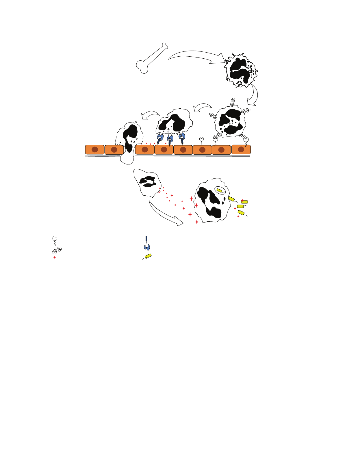
tides (Figure 1). Upon encountering bacteria neutrophils
monomers to filamentous F-actin polymers [3]. Spontaneous
capture, ingest, and kill them by production of reactive
nucleation of actin filaments is slow since, unlike the polymer
oxygen species. Abnormalities in any aspects of neutrophil
which is stabilized by contacts between several subunits,
development and/or function induce immunodeficiency or
dimers and trimmers are unstable. Cells control new filament
aberrant inflammatory reactions (Table 1) which reflects in
assembly through the induction of nucleation promoting
the complexity of the diagnosis of these diseases [2]. A
factors such as the WASp/WAVE (Wiskott-Aldrich syndrome
common denominator in these diseases is failure to properly
protein/WASp-family verprolin-homologous protein) family
regulate the actin cytoskeleton by direct or indirect genetic
proteins. These factors stimulate the Arp2/3 protein complex
mutations. Such failure is implicated in decreased migratory
to nucleate actin polymerization in the side of an existing
and adhesive properties, altered vesicle dynamics and release,
polymer as a branch. New filaments grow rapidly, in a rate
and perturbed assembly of the NADPH oxidase necessary
limited by the concentration of available actin monomers,
for antimicrobial killing by neutrophils. Here we propose
and they push the plasma membrane forward. This transient
that the failure to regulate the actin cytoskeleton and vesicle
growth is terminated by the binding of capping proteins 2 Journal of Immunology Research
Myeloid hematopoiesis, neutrophil apoptosis
SCN1 (ELANE), SCN2 (GFI1), SCN3 (HAX1),
SCN4 (G6PC3), SCN5 (VPS45) XLN (WASp) Egress from the bone marrow 𝛽-Actin WHIM syndrome (CXCR4) Cohen (VPS13B)
P14-deficiency (MAPBPIP) CMTDIB (DNM2)
Hermansky-Pudlak2 (AP3B1) Firm adhesion Rolling Chédiak-Higashi (LYST) LAD I (𝛽2 integrins) LAD II (SLC35C1) Griscelli2 (RAB27A) LAD III (FERMT3) XLN (WASp) SDS (SBDS) XLN (WASp)? GSD1B (G6PT1) Activated endothelium Chemotaxis, migration Phagocytosis, superoxide
Neutrophil immunodeficiency (Rac2) Neutrophil XLN (WASp)? Immunodeficiency (Rac2)
NAD (?), NAD47/89 (LSP1) CGD (PHOX proteins) SCN3 (HAX1)? SCN4 (G6PC3) SCN4 (G6PC3) XLN (WASp)? SCN5 (VPS45) NAD (?) 𝛽-Actin NAD47/89 (LSP1) Chédiak-Higashi (LYST) 𝛽-Actin PLS (CTSC) GSD1B (G6PT1) SDS (SBDS) SCN5(VPS45) CGD (PHOX proteins) GSD1B (G6PT1) Integrin ligand Selectin S-Lex Integrin Chemotactic molecule Bacteria
Figure 1: Neutrophil development, migration, and function. Diseases described in the review are indicated where they are believed to act.
to the fast-growing (barbed) end of the filament. Breakage
2.1. 𝛽-Actin Dysfunctions. Actin proteins are highly con-
of filaments is catalyzed by actin-severing proteins such
served evolutionary in vertebrates and their functional
as gelsolins and the ADF/cofilin family. Severed filaments
integrity is essential for the survival of a complex organism.
shorten and debranch. As a result of the action of nucleation
Out of the six actin isoforms, the nonmuscle 𝛽-actin is
promoting factors, capping and actin severing proteins, and
ubiquitously expressed in all cell types and the deletion of this
several other actin binding accessory proteins, F-actin forms
isoform is embryonic lethal in mice [5, 6]. A single case study
a tightly regulated 3-dimensional network which is growing
of a patient carrying a heterozygous 𝛽-actin E364K mutation
in the leading edge and disassembles some distance in the rear
reported recurrent infections, thrombocytopenia, photosen-
thereby creating a plasma membrane protrusion [3].
sitivity, and mental retardation [7]. The patient exhibited
Neutrophils polarize their cytoskeleton to form a leading
profound neutrophil functional defects in chemotaxis, super-
edge (lamellipodia or pseudopod) towards the signal of origin
oxide production, and membrane potential response. These
and a trailing uropod in the posterior of the cell. While the
defects were attributed to impaired binding of the E364K 𝛽-
leading edge consists of highly branched and dynamic actin
actin to the actin-binding protein, profilin. Another mutation
filaments, the uropod is rich in actin-myosin II contractile
in 𝛽-actin, R183W, causes malformations, deafness, and
structure. During chemotaxis, the cells extend the leading
neurological abnormalities such as dystonia [8]. Yet another
edge by local actin polymerization and contract the uropod
set of mutations in 𝛽-actin have been recently identified
to allow movement in the direction of the signal. It has
to cause Baraitser-Winter syndrome (BRWS). BRWS is a
become increasingly clear that the Rho GTPases Cdc42, Rac,
rare condition, characterized by ocular colobomata, ptosis,
and Rho serve a key role in establishment of cell polarity.
neuronal migration defect, distinct craniofacial anomalies,
By direct binding to the WASp family of proteins they
and intellectual disability [9–11]. Remarkably, the neutrophil
regulate localized actin polymerization and interaction with
dysfunction (𝛽-actin E364K), dystonia (𝛽-actin R183W) [8],
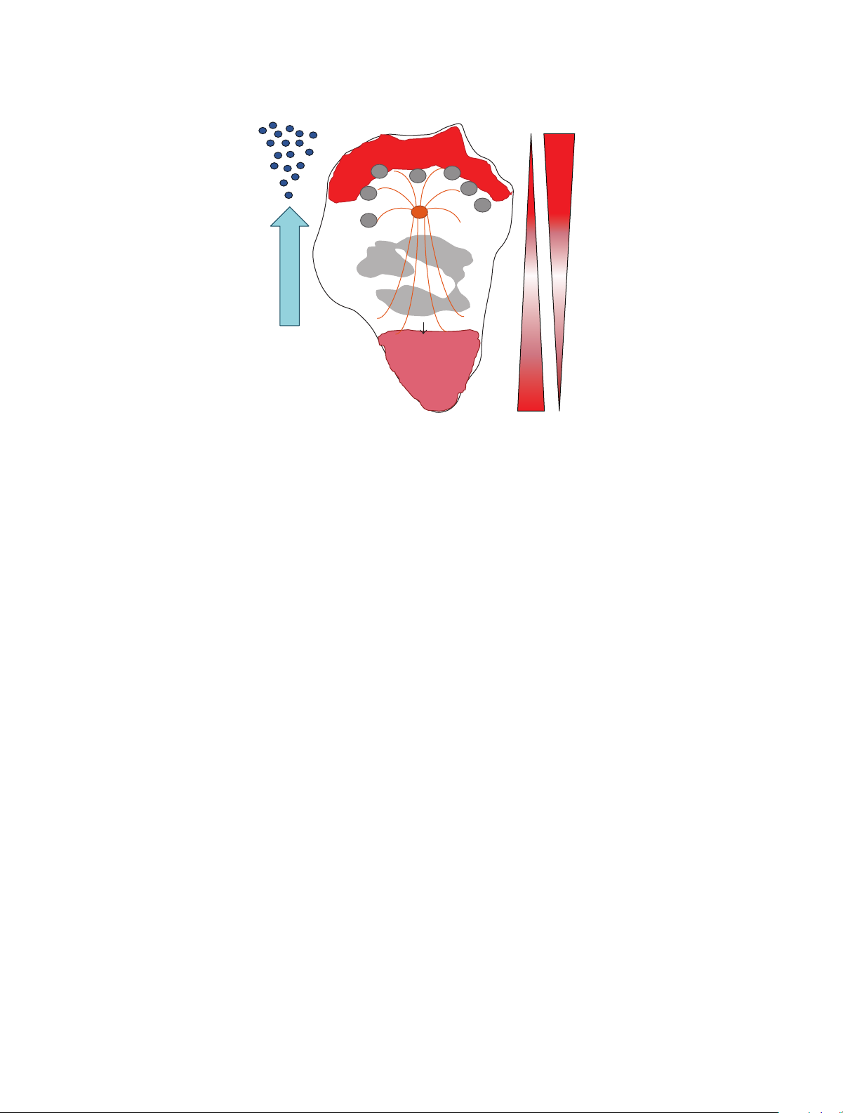
cell surface chemokine receptors and integrins [4] (Figure 2).
and BRWS [7, 9] cases were presumably caused by dominant Journal of Immunology Research 3 s n ia, tal re, n g ia atio en en en p n ts sis statu atio p - est enia o o linemia yto leedin re,m atio p h u logical men yto rt tard b p yp b anif tu ho h p o ir oc sho d m boc tard pa III: ym glo m sta uk re ym eur II: talre D an ro rt L N im Le en elll ther o D A mma A m L O Th sh L Bc ga s s s n n us K P n s 4 S 4X and er u tio tatio 70 72P 76 h of u 36 tatio E L2 S2 I2 I294T D57N W4 2%) ot vario ermin M (7 mu runca T C-t itance D L R R D A X AD AR AR A A A her In s n + + + + + + fectio In osis cyt o + + + mics. Phag yna d il h xide p ero +++ +++ + tro p Su neu in n ts ec hesio + + + + ef d A tald geni taxis n o + + + + ++ emo h 1:C C ble a T enia p + +? ++ tro eu N n/ o sis) n o elet esio opt h arget skeleton osk p T to yt A llad Cy (C Ce 𝛽2 1 tin 1 in y Sp I: II: III: ene ? P1 mil D 35C D CR4 G 𝛽-Ac WA Rac2 LS HAX D tegr fa A C A CX LA in L SL L FERMT3 n tin tio enia y n D) e/ 𝛽-ac unc p A tal esio D) me f eas (SCN3) o o ith ysf tro (N /89 3 A n w d n dis geni adh (L il neu il eficienc ilactin 47 n ndr h h d h tio D enia y sy e p me p nn co yte tatio ated p p ked A uno o oc eas u ci tro tro tro unc N ma tro m o u ndr u st ysf d o uk Dis A ass neu X-lin Ne imm sy Ne d an K Severe neu Le deficienc WHIM 4 Journal of Immunology Research s n , , , , , n y, n y, n y, n y d sis ic sis ic sis n e rt an ro atio y, ly ects sis s logic tatio cyt cyto tatio cyt cyto tatio re tatio atio fib ti ic to est ess y hal enia asis ti en nia def y,sho athies, io en io en en et, eficienc h w p efects, glucos n p eficienc eficienc eficienc statu d p tard to mega d ost o ects anif o d d d cep o o ho tal phago hist phago hist creat rt atel eakn ro o rro p ed od n m igm igm igm igm p p p p pl w at yp art eni ir me erkerato def o eur uno ho o uno ho o uno o uno talre icr h ma p eri Pa e,hema p b ym pa N p p p p sho p M nephr L He og ho P sufficienc ther hemo hemo m en ne Hy o ur Im atur O Hy imm lym Hy imm lym Hy imm Hy imm Li M in B st s n s s s s s s s s s s s u u u u u N 4 u u u u u u 82S tatio 38K 03R rio u vario vario 3’UTR vario vario vario T22 E2 vario N3 K4 vario vario va vario vario M itance R R R D R R D D R R R R A A AR A A A A A A A XL AR AR AR AR A A her In s n + + + + + + + + fectio In osis cyt o Phag xide . ero + + + ed p u Su tin n n o 1:C hesio d le A b Ta taxis ++ ++ emo h C enia p ++ ++ + ++ +A ++ ++ ++ ++ ++ ++ ++ tro eu N . ed lar rt, o g -link X sp enesis, er arget sicu n rtin T iog XL: Ve tra b so Oth t, n ina m o d P 𝑥 𝑥 PI 2 𝑥 𝑥 𝑥 S 𝑝ℎ𝑜 𝑝ℎ𝑜 mal ene ST 27A SC B M 𝑝ℎ𝑜 PB ANE 𝑝ℎ𝑜 𝑝ℎ𝑜 D so G LY A GFI1 91 22 CT SB to RA AP3B1 DN VPS13B VPS45 EL G6PC3 G6PT1 𝑝47 𝑝67 𝑝40 M 𝑔𝑝 𝑔𝑝 au e s u d typ th n e o D1B) ato o ve,AD: t (SCN5) m lak o n e m m o d 2 T ; n tal tal tal (SCN4) (GS e y u e B m (SCN2) o (SCN1) 1 2 4 rage 1b ulo DS) recessi igashi n `evr typ arie- mina tatio geni geni a geni e f GD) (PLS) n-Dia (S syndr u sto sky-P iate IB) n n n enia eni enia gra -Le mal e k-H me me t-M syndr m co co co typ (C n me ma me so o an o e,do ed 5 p p p h 4 e nic e o o o to eas celli ´edia rm tro tro tro ill au ndr arco ro ris 4-deficienc rm ndr eas hen eas eas h te o h ap ndr wac ndr Dis Ch sy G 2 P1 He sy C dis in (CMTD C VPS Severe neu Severe neu Severe neu Glycogen dis C dis P sy Sh sy AR: Journal of Immunology Research 5 Chemotactic molecule Actin Lamellipodia dynamics Cortical actin Rac2 Cdc42 MTOC microtubule Cdc42? Migration WASp direction 𝛽2 integrins actin-myosin RhoA Uropod Actin stability
Figure 2: Neutrophil polarity during migration. The role of the cell cytoskeleton and the proteins that regulate cell polarity is indicated.
missense mutations in 𝛽-actin. Although no immunological
in all responses that depend on the actin cytoskeleton such as
defects were reported either in the 𝛽-actin R183W case or
F-actin polymerization, migration, adhesion under flow, and
BRWS cases, both reports found abnormal F-actin structures
𝛽2-integrin clustering [14, 15]. WASp−/− neutrophils exhibit
in mutant 𝛽-actin transfected cell lines [8, 9]. The BRWS
multiple F-actin fronts and fail to redistribute CD11b into
associated R196H mutation induces greatly increased F-
clusters at the uropod [14, 16]. A recent report shows that
actin with multiple, anomalous F-actin-rich, filopodia-like
in neutrophils, WASp seems to be dispensable for F-actin
protrusions compared to control cells in lymphoblastoid cell
polymerization at the leading edge [16]. Instead, Cdc42
lines [9]. Both the BRWS mutation R196H and the dystonia
activates WASp at the uropod and facilitates microtubule
mutation R183W mutation render F-actin more resistant to
capture and stability at the uropod via clustering of CD11b
the depolymerizing effect of Latrunculin A in lymphoblasts. 𝛽2 integrins [16].
These results suggest that accumulation of filamentous actin
The more recently described X-linked neutropenia (XLN)
plays an important role in diseases caused by mutations
is caused by mutations (L270P, S272P, I276S, and I294T)
in 𝛽-actin. While there is yet no evidence that the R183W
in the GTPase binding domain of WASp and destroys
and BRWS mutations in 𝛽-actin affect the immune system
the autoinhibited conformation of WASp [12, 13]. These
broadly, given the neutrophil dysfunction in the E364K
mutations were initially predicted to lead to constitutively
patient together with the central role and abundance of 𝛽-
active WASp and as a consequence cells would have increased
actin in leukocytes, we reason that neutrophil function is
load of polymerized actin [17]. Several laboratories have now likely to be compromised.
confirmed this hypothesis and shown markedly increased
polymerized actin in neutrophils, in macrophages, and in
2.2. WASp Deficiency and Overactivity. Patients with
B and T cells [18–22] (Keszei and Westerberg-unpublished
Wiskott-Aldrich syndrome (WAS) lack or have reduced
observation). XLN patients suffer from recurrent bacterial
expression of WASp and suffer from combined immu-
infections because of severe neutropenia and monocytopenia
nodeficiency with recurrent infections [12, 13]. WASp is
[17, 18, 20] and they may develop cytogenetic changes
uniquely expressed in hematopoietic cells and resides as
indicative of chromosomal instability, myelodysplasia, or
an inactive form in the cytoplasm due to an autoinhibited
acute myeloid leukemia [18–20, 22]. Neutrophils from XLN
folding where its GTPase binding domain forms a molecular
patients have decreased capacity to phagocytose bacteria and
interaction with the carboxy-terminal verprolin-cofilin
kill them [18]. Oxidative burst in XLN neutrophils is normal
homology and acidic (VCA) domain. Upon signaling, the
in response to PMA, while receptor-mediated oxidative burst
small Rho GTPase Cdc42 binds to WASp that undergoes a
in response to E. coli or fMLP is reduced [18]. This suggests
conformational change to open up the protein. This exposes
that XLN neutrophils fail to effectively assemble signaling
the carboxy-terminal part of the protein that binds directly to
complexes at the cell membrane. One recent report shows
the Arp2/3 complex and induces actin polymerization. It may
that excess cytoplasmic F-actin in XLN causes increased
not be surprising that neutrophils lacking WASp have defects
cellular viscosity and tension and this indirectly perturbed 6 Journal of Immunology Research
mitotic mechanics [23]. Membrane tension appears to be
the NADPH oxidase, including gp91phox (cytochrome b-
one mode of long-range inhibition mechanisms. Membrane
245, 𝛽-polypeptide, CYBB), p22phox (cytochrome b-245, 𝛼-
tension nearly doubles during leading edge protrusions, and
polypeptide, CYBA), p47phox (neutrophil cytosolic factor
increase in tension is sufficient for long-range inhibition
1, NCF1), p67phox (NCF2), and p40phox (NCF4). CGD
of Rac activation at the leading edge [24]. In contrast,
patients have defective microbial activity resulting from
reduced membrane tension activates actin assembly through-
abolished superoxide production. Studies of CGD patients
out the cell [24]. Macrophages from XLN patients have
neutrophils suggest that assembly of the NADPH complex
increased turnover rate of actin-rich adhesive structures
is not only important for oxidative killing of microbes. The
called podosomes [18] and murine XLN B and T cells can
microbial spectrum of infections in CGD includes bacteria
adhere to antibody-coated layers but fail to coordinate cell
that require neutral pH for effective nonoxidative killing and
spreading [22]. B cells from XLN patients form less dynamic
are resistant at the acid pH found in the phagosomes of
contacts with L-selectin ligands under flow [21]. This is
CGD neutrophils. These include S. aureus, S. marcescens,
likely to be caused by excessive localized production of
N. asteroids, and A. fumigatus. This implies that reactive
cortical F-actin that induces increased rigidity of microvilli
oxygen species produced by the NADPH oxidase also act as
[21]. Neutrophils devoid of Rac2 (discussed below) are also
intracellular signalling molecules, leading to the activation
unable to adhere to L-selectin ligand under flow despite
of other nonoxidative pathways for microbial killing. One
normal levels of L-selectin expression [25]. Together this
possible mechanism whereby reactive oxygen species could
highlights the importance for dynamic cytoskeletal rear-
contribute to lamellipodia and thereby increased motility
rangement in L-selectin-dependent rolling on endothelial
of neutrophils is through cofilin. Reactive oxygen species
cells. How increased load of polymerized actin in XLN affects
induce cofilin dephosphorylation through activation of the
cell polarity, migration, and tension in neutrophils remains to
cofilin phosphatase Slingshot [32]. When dephosphorylated, be determined.
cofilin binds existing cortical actin filaments and severs them.
This generates new barbed ends on the filaments to which
the Arp2/3 complex can bind and stimulate branching and
2.3. Neutrophil Immunodeficiency Syndrome (Rac2). Rac2
thereby increase dynamics of the lamellipodia [33]. One
belong to the Rho family of small GTPases that act as
implication is that, in the absence of NADPH oxidase activity,
molecular switches inside the cell by cycling between a GDP-
neutrophils have less capacity to form a dynamic lamellipodia
bound inactive form and a GTP-bound active form [26]. The
required for migration [34] and that phagocyte enzymes are
activity of Rho GTPases also depends on their localization
present but hypofunctional [35].
to lipid membranes by posttranslational addition of lipid
anchors. In neutrophils, Rac2 is highly polarized to the
leading edge where it regulates actin assembly by activating
2.4. Neutrophil Actin Dysfunction (NAD) Syndrome. One
the WASp family members. Another Rho GTPase, RhoA, is
case of neutrophil actin dysfunction (NAD) was reported
localized to the trailing uropod where it coordinates actin-
in 1974 in a male newborn patient [36]. The patient had
myosin filaments. A third Rho GTPase member, Cdc42, is a
recurrent bacterial infections despite marked neutrophilic
key regulator of cell polarity by assembly of the microtubule
leukocytosis, impaired neutrophil migration from blood to
organizing center (MTOC) between the leading edge and the
the inflammation site, and impaired phagocytosis by neu-
cell nucleus. Rac2 is highly expressed in neutrophils and is
trophils. The patient’s neutrophils extended a few fork-like
essential to assembly of the NADPH oxidase that initiates
pseudopodia and actin isolated from his neutrophils poly-
production of toxic oxygen metabolites to kill pathogens [27].
merized poorly in vitro. F-actin content in the neutrophils of
Three patients with mutations in Rac2 have been identified
the patient’s father, mother, and sister was significantly lower
that suffer from a neutrophil immunodeficiency syndrome.
than in controls [37]. Expression of CR3 subunits (CD11b,
Curiously, all three patients harbor a D57N mutation within
CD18) was depressed in the patient’s mother and a sister,
the DX2G motif, conserved in all GTPases, that results in
which argues that NAD is a form of leukocyte adhesion
a dominant negative protein. Rac2-D57N neutrophils show
deficiency (LAD, discussed below); however, F-actin content
complete loss of chemotaxis, azurophil granule secretion,
is normal in LAD patients [38]. It had been speculated that
superoxide generation, and polarization in response to a
NAD is a result of a defect in an actin associated protein;
variety of receptor stimuli, especially the chemokine fMLP
however the gene mutation which caused NAD in the index
[28–30]. Murine Rac2−/− neutrophils show a similar pheno- patient had not been found.
type and have perturbed polarization and decreased capacity
Defective actin polymerization was also found in a 2-
to migrate in vitro and in vivo into the peritoneum [25].
month-old male infant with recurrent fevers and fungal
Moreover, Rac2−/− neutrophil have decreased NADPH func-
infections [39]. The neutrophils of the patient had fre-
tion associated with reduced clearance of the opportunistic
quent development of F-actin rich filamentous projec- pathogen A. fumigatus.
tions that were not present in control PMNs and showed
The critical role of NADPH activity for neutrophil
profound defect in random migration, chemotaxis toward
function is highlighted in chronic granulomatous disease
fMLP, and phagocytosis. In this patient, CD11b expression
(CGD), characterized by severe, life-threatening bacterial
was increased. In contrast to the other NAD case, cell
and fungal infections and immune dysregulation [31]. CGD
lysates from this patient showed a significant decrease in
is caused by mutation in any one of the five subunits of
an 89 kDa protein and a marked increase in a 47 kDa Journal of Immunology Research 7
protein. Coates and colleagues named this disease actin
been shown in knock-out mouse studies that CD11b cluster-
dysfunction NAD 47/89. The overexpressed 47 kDa protein
ing is abrogated in WASp and Cdc42 deficient neutrophils
has been shown to bind actin and its cloning revealed that
[16] and the Cdc42/WASp axis acts upstream of integrin
it was a known actin regulator, lymphocyte-specific protein
functions. These studies suggest that WASp might regulate
1 (LSP1) [40], which is expressed in normal neutrophils [41].
inside-out integrin signaling in neutrophils and it is critical
LSP1 overexpression produces F-actin bundles and hair-like
to maintain neutrophil polarity during migration [16].
surface projections in several eukaryotic cell lines. More-
over, increased expression of LSP1 inhibits the locomotion
2.6. Hax1 Deficiency. Approximately 15% of severe congenital
of normally motile human melanoma cells [42]. On the
neutropenias (SCNs) are caused by autosomal recessive
other hand, murine neutrophils devoid of LSP1 expression
mutations in the HAX1 gene [48, 49]. Patients with HAX1
have increased migratory capacity. Together these data show
mutations present marked neutropenia (absolute neutrophil
that LSP1 is a negative regulator of neutrophil chemotaxis
count < 500 𝜇L−1) which causes life-threatening bacterial [43].
infections in newborns. HAX1 is involved in B-cell receptor
signaling [50] and it has been shown to regulate apoptosis
2.5. Leukocyte Adhesion Deficiency (LAD). During the course
[51, 52]. Neutrophils from HAX1-deficient patients showed
of an infection neutrophils leave the blood stream in large
higher rate of spontaneous and TNF𝛼 induced apoptosis than
numbers by transmigrating the endothelium. The complex
control neutrophils due to loss of mitochondrial membrane
process of transmigration is tightly regulated in order to
potential. It has been suggested that HAX1 is a major inhibitor
segregate the homeostatic tissue environment from blood
of apoptosis in myeloid cells and that neutropenia in HAX1-
vessels which carry a large number of potentially damaging
deficient SCN patients is caused by lack of this antiapoptotic
leukocytes. Local inflammation quickly activates the adjacent
function [49]. HAX1 has been shown to interact directly
endothelium which upregulates P- and E-selectins that binds
with adhesion and cytoskeleton regulating proteins, such as
to sialyl-LewisX carbohydrates on the neutrophil surface.
the actin nucleation-promoting factors cortactin [53] and
Swiftly moving neutrophils in blood vessels get tethered to the
its homolog hematopoietic lineage cell-specific protein 1
endothelial surface by selectins and they start rolling on that
(HS1) [50], 𝛽6 integrin [54], and G𝛼13 [55]. Cavnar and
surface. Chemoattractants, such as CXCL8 (IL-8), activate
colleagues demonstrated that Hax1 predominantly localize in
𝛽2 integrins on neutrophils which in turn bind intercellular
the leading edge in the PLB-985 neutrophil-like cell line [56].
adhesion molecule-1 and molecule-2 (ICAM-1, ICAM-2)
Knock-down of HAX1 expression results in impaired motility
on the activated endothelium and mediate firm adhesion
and elongated uropods, as well as decreased RhoA activity.
between neutrophils and the endothelium. This firm adhe-
Impaired uropod detachment in HAX1-deficient neutrophils
sion is prerequisite for extravasation. Aberrations in these
is caused by increased integrin mediated adhesion similarly to
processes in LAD patients lead to recurrent skin infections
neutrophils devoid of RhoA expression. The authors suggest
and soft tissue abscesses, periodontal disease, and impaired
that HAX1 is a negative regulator of integrin-mediated adhe-
pus formation despite blood neutrophilia [44]. While LAD II
sion in neutrophils by affecting Rho GTPase signaling [56].
is a result of mutations in a membrane transporter of fucose
which impairs selectin mediated adhesion, LAD I is caused by
2.7. WHIM Syndrome. Warts, hypogammaglobulinemia,
a genetic defect in CD18 (ITGB2). CD18 is a common 𝛽 chain
infections, and myelokathexis (WHIM) is an immuno-
of four 𝛽2 integrins in leukocytes, each containing a different
deficiency with autosomal dominant inheritance. In most 𝛼 chain: LFA-1 (𝛼 𝛽 𝛽
L 2 or CD11a : CD18), Mac-1 (𝛼M 2 or
kindred gain of function mutations of the chemokine
CD11b : CD18 which is complement receptor CR3), gp150/95
receptor CXCR4 have been identified as the cause of the (𝛼 𝛽
X 2 or CD11c : CD18 which is complement receptor CR4),
disease [57]. CXCR4 on neutrophils and its ligand, stromal and ADB2 (𝛼 𝛽
D 2 or CD11d : CD18). Mutations in CD18 fully
cell-derived factor 1 (SDF1; also known as CXCL12) in the
or partially abolish the expression of 𝛽2 integrins on leukocyte
bone marrow stroma, are major bone marrow retention
surface, thereby largely impeding neutrophil transmigration
factors for neutrophils [58, 59]. According to a current
into inflamed tissues and renders neutrophils unresponsive
hypothesis, increased CXCR4-mediated retention signals in
to bacteria opsonized with complement fragment C3bi. In
bone marrow lead to myelokathexis (hyperplasia with an
contrast, LAD III patients show normal expression of 𝛽2 inte-
accumulation of apoptotic neutrophils in the bone marrow)
grins. Due to mutations in the intracellular protein kindlin-
and neutropenia in the periphery [60].
3 (FERMT3) which regulates inside-out integrin activation,
Various early stop codon mutations in WHIM patients
the integrins fail to change their conformation to become
have been identified to cause C-terminal intracellular trun- functionally active.
cations in the CXCR4 protein [57, 61]. Accumulating
Integrins clearly depend on the connection to the actin
evidence shows that C-terminal truncations in CXCR4
cytoskeleton to carry out their functions [45–47]. They bind
impair ligand-induced desensitization and internalization
to several F-actin associated proteins (talin, vinculin, and
of CXCR4. Thereby, an important physiological negative
𝛼-actinin) [46]. Besides anchoring themselves to the actin
feedback mechanism is interrupted in which CXCR4 activ-
cytoskeleton, integrins are also involved in induction of local
ity is downregulated to release neutrophils from the bone
actin polymerization where they engage their ligands on the
marrow [60–63]. Intriguingly, WHIM transgenic zebrafish
extracellular matrix on other cells [46]. Intriguingly, it has
neutrophils show prominent random membrane protrusions 8 Journal of Immunology Research
but impaired persistent motility in vivo which resulted in
reduced bactericidal activity [72]. A key feature of CHS is
neutrophil retention within areas of high SDF1𝛼 expression.
the presence of giant granules in most nucleated cells due
to aberrant vesicle fusion or fission. Neutrophil granules are
deficient in cathepsin G and NE [73] and mobilization of
3. Defects of Vesicular Transport
the giant granules is impaired in CHS patients [74]. In fact,
enlarged granules might impair cell kinetics mechanically
Neutrophils kill microbes by controlled release of microbici-
[71]. Mutations in LYST, a lysosomal trafficking regulator
dal products from their secretory granules to the extracellular
gene, have been identified as the cause of CHS [75].
space and by elimination in neutrophil phagosomes. Neu-
Griscelli syndrome type 2 is characterized by partial
trophils contain four types of secretory organelles: primary
albinism and marked immunodeficiency including frequent
(azurophil) granules, secondary (specific) granules, tertiary
pyogenic infections associated with neutropenia [76]. Muta-
(gelatinase) granules, and secretory vesicles. Out of the four
tions in the small GTPase RAB27A gene were identified as
organelles, secretory vesicles are mobilized readily, probably
the cause of disease [77]. The Rab family of GTPases control
already during neutrophil rolling on activated endothelia,
trafficking of vesicles between intracellular compartments to
and they carry membrane associated proteins such as the 𝛽2
target membranes. Studies on mutant and gene targeted mice
integrin component CD11b to the plasma membrane. This
suggest that Rab27a controls exocytosis of azurophil vesicles
process is thought to transform circulating neutrophils into in neutrophils [78–80].
a highly responsive cell, primed for migration [64]. Gelati-
p14 deficiency was described by Bohn and colleagues
nase granules and specific granules are mobilized next and
in 2006 [81]. Four out of 15 offspring in the index family
they carry, among other effectors, gelatinase and lactoferrin,
developed recurrent bronchopulmonary infections, hypopig-
respectively. Azurophil granules need the strongest stimulus
mented skin, and neutropenia. The clinical phenotype of
for their release and they mainly contain myeloperoxidase
p14 deficiency was unique among the other described
(MPO), defensins, and neutrophil elastase (NE). Regulated
hypopigmentation-associated immunodeficiencies by caus-
secretion of granules in neutrophils is a complex process
ing short stature in the affected individuals. In vitro exper-
which requires sorting of the proteins to this pathways,
iments showed impaired bactericidal activity and abnormal
guiding transport vesicles specifically to secretory granules
azurophil granules in p14 patient neutrophils. Furthermore,
and mediating membrane fusion and fission. Moreover,
the distribution of the late endosomal compartment is
vesicle trafficking critically relies on the interplay between the
perturbed in the absence of p14. The p14 protein is an
microtubule and actin cytoskeleton. Among others, the small
adaptor of the MP1-MAPK scaffold complex and is involved
GTPase Cdc42 has the capacity to link these two molecular
in localization of MP1-MAPK to endosomes. The authors
motor systems to maintain cell polarity. Cdc42 coordinates
suggest that p14 is involved in granulocyte colony-stimulating
the microtubule cytoskeleton by binding to the Cdc42 inter-
factor (G-CSF) receptor signaling.
acting protein (CIP4) that directly regulates microtubule
assembly and induces membrane deformation [65]. Cdc42
also coordinates actin polymerization via the activation of
3.2. Mutations in Neutrophil Elastase and AP3. More than
WASp and its relative the neuronal (N)-WASp that upon
50% of patients with congenital severe neutropenia and
Cdc42 binding becomes active and induces actin polymeriza-
nearly all patients with cyclic neutropenia harbour mutations
tion via the Arp2/3 complex [66, 67]. In this way, Cdc42 can
in the ELANE gene encoding for the neutrophil elastase (NE),
mediate the interaction between actin and microtubules and
a broad-specificity serine protease localized in azurophil
regulate vesicle trafficking. Since neutrophils are packed with
granules [82–84]. The mechanism for how autosomal domi-
potentially harmful substances in granules, correct sorting
nant mutations in ELANE induce neutropenia is still unclear
and release of vesicles is key for neutrophil survival and
[85]. The known human mutations do rarely affect protease
function. It is reasonable to predict that any change in vesicle
activity of NE, nor its properties for substrate specificity [83].
trafficking or localization of vesicle components would be
Once produced, NE binds the adaptor protein 3 (AP3) and harmful for the neutrophil.
is shuttled from the trans-Golgi to azurophil granules. It is
possible that ELANE mutations lead to mislocalization of NE
3.1. Neutropenias with Hypopigmentation. The function of
within the cell or disturb NE protein folding [86]. Disruption
neutrophils, cytotoxic T lymphocytes, natural killer cells,
of either NE or its cargo protein, the lysosomal transporter
and mast cells is highly dependent on intact secretory
AP3 (encoded by AP3B1) [87, 88], perturbs the intracellular
machinery for the capacity of these cells to degranulate
trafficking of NE to azurophil granules [89]. Moreover,
and release vesicular content towards pathogens and target
mutated NE can induce the unfolded protein response in
cells. Genetic defects in degranulation often coincide with
the endoplasmic reticulum [90, 91]. A recent report shows
impaired melanin secretion by melanocytes indicating the
that certain patient mutations in ELANE force transcription
usage of similar secretory pathways [68].
to an alternative start site in the gene and production of an
Ch´ediak-Higashi syndrome (CHS) is characterized by
amino-terminal truncated form of NE that lack ER-localizing immunodeficiency, hypopigmentation, and neurologic
(pre) and zymogen-maintaining (pro) sequences yet retain
symptoms [69]. Patients develop recurrent pyogenic infec-
essential catalytic residues [85]. The key role of ELANE in
tions and often periodontal disease which is associated with
neutrophil homeostasis is also indicated by the development
neutropenia [70], impaired neutrophil chemotaxis [71], and
of SCN in patients carrying dominant negative mutations in Journal of Immunology Research 9
the GFI1 gene,which is a transcriptional repressor of ELANE
and G6PC3 deficient neutrophils are impaired in chemotaxis,
[92]. Although the mechanism for SCN induced by ELANE
respiratory burst, and calcium mobilization [101, 102].
mutations is not directly linked to the actin cytoskeleton,
Papillon-Lef`evre syndrome (PLS) is characterized by
it is likely that neutrophil deficiency that affects the actin
palmoplantar keratosis and severe periodontitis which results
cytoskeleton may have similar mislocalization of neutrophil
in premature tooth loss [105]. PLS is caused by mutations
proteases to vesicles and/or activation of the unfolded protein
in cathepsin C (CTSC) [105, 106], a lysosomal protease response.
which is expressed highly in epithelial cells [106] and
immune cells, including polymorphonuclear cells [107] and
alveolar macrophages. In immune cells, cleavage by CTSC
3.3. Other Neutropenias with Vesicle Sorting Defects. Charcot-
activates a variety of granule serine proteases by removing
Marie-Tooth disease (CMT) is a progressive disorder of the
their inhibitory N-terminal dipeptides. Among others, CTSC
peripheral nervous system and a genetic variant of CMT is
targets are the neutrophil effectors NE, cathepsin G, and
caused by mutations in dynamin-2 (DNM2) [93]. DNM2
proteinase-3 [108, 109]. Increased susceptibility to infections
is a ubiquitously expressed mechanochemical protein with
in some cases [110] and neutrophil chemotaxis deficiency
GTPase activity. DNM2 is associated with microtubules and
was reported in PLS patients [111]. It is controversial whether
is involved in endocytosis, cell motility, and centrosome
neutrophil chemotaxis is intrinsically defective in CTSC-
organization. Several CMT patients with K558E and K558del
deficient neutrophils. Based on the CTSC (also called dipep-
DNM2 mutations have neutropenia [93]. The mechanism
tidyl peptidase I; DPPI) knock-out mouse model, Adkison
how DNM2 mutations cause neutropenia is unknown.
and colleagues argue that neutrophil-derived serine proteases
Cohen syndrome is a multiple congenital anomalies-
are involved in the regulation of cytokine production at sites
mental retardation syndrome which is associated with neu- of inflammation [109].
tropenia [94, 95]. No bone marrow morphological abnormal-
Shwachman-Diamond syndrome (SDS) is characterized
ities were observed in Cohen syndrome patients; however
by pancreatic insufficiency, pancytopenia, and leukemia
their neutrophils exhibited greater adhesive capacity than
predisposition [112]. Bone marrow failure in patients with
the control ones and CD11b and CD62L surface expression
SDS is often manifested in neutropenia and peripheral
was decreased on their neutrophils [96]. Cohen syndrome
SDS neutrophils are defective in chemotaxis towards fMLP
is caused by mutations in the vacuolar protein sorting 13B
[113, 114]. This disease is caused by mutations in the SBDS
(VPS13B) gene [97]. Although the exact pathomechanism is
gene, encoding for a predicted RNA-processing protein, and
unknown, vacuolar sorting proteins are involved in endo-
suggests that SDS may be involved in RNA metabolism [115].
somal trafficking and protein recycling in the trans-Golgi
Even the most common genetic disease Chromosome
network. Indicating their importance in granulocyte devel-
21 trisomy or Down syndrome causes a wide range of
opment, another VPS protein, VPS45 was recently found to
mild primary and secondary immunodeficiencies related to
be mutated in severe congenital neutropenia patients [98, 99].
neutrophil dysfunction [116]. Trisomy 21 is characterized by
In accordance with other severe congenital neutropenias,
high frequency of infections in the upper respiratory tract
VPS45 mutant patients had severe infections and their neu-
and periodontal disease which at least partially is attributed
trophils and bone marrow myeloid cells showed accelerated
to reduced neutrophil chemotaxis [117].
apoptosis. Peripheral neutrophils showed impaired migration
and impaired superoxide production [98]. Vps45 is a member
of the Sec1/Munc18 protein family that regulates the assembly
of specific SNARE complexes. SNARE proteins mediate the
5. Conclusion and Perspective
fusion of lipid bilayers and serve a vital role in homeostasis of
The dynamics of the actin cytoskeleton is a key feature
vesicle transport within the cell.
of rapidly moving and acting cells such as neutrophils. A
striking feature of neutrophil deficiency is that of all the
4. Other Neutrophil Deficiencies with
hematopoietic cells, neutrophils are exceedingly vulnerable Chemotaxis Involvement
to loss of specific proteins or to changes in their activity.
The reasons of this vulnerability perhaps originate from their
Severe congenital neutropenia 4 (SCN4) is caused by
unique developmental and functional requirements.
homozygous mutations in the ubiquitously expressed cat-
Neutrophils have a high turnover rate; they live for an
alytic subunit 3 of the glucose-6-phosphatase gene (G6PC3)
average of 5 days in man [118] with a half-life of 7–10 hrs
[100]. Besides recurrent bacterial infections and neutropenia,
in human circulation [119]. A vast output of 1011 mature
SCN4 patients also show structural heart defects and uro-
neutrophils/day from bone marrow requires efficient cell pro-
genital abnormalities. Importantly, neutrophil development
liferation in the myeloid lineage, terminal differentiation, and
and function is also severely impaired in glycogen storage
egress from bone marrow. Defects in any of these processes
disease type Ib (GSD-Ib) which is caused by mutations in the
cause SCN. An archetype of actin cytoskeleton disease that
glucose-6-phosphate transporter 1 (G6PT1) [101, 102]. Chou
results in SCN is XLN, caused by overactivity of WASp. Given
and colleagues argue that a glucose-6-phosphatase complex
that all hematopoietic cells are dependent on WASp for their
which is composed of G6PC3 and G6PT1 is essential for neu-
function it is reasonable to predict and evidence suggests
trophil energy homeostasis and functionality by regulating
that increased load of polymerized actin in XLN would affect
endoplasmic reticulum glucose storage [103, 104]. Both G6PT
the immune system broadly [18, 19, 21, 22]. However, the 10 Journal of Immunology Research
cardinal clinical feature of XLN patients is still neutropenia
74/89, or in actin itself as in 𝛽actin deficiency. Moreover,
and neutrophil dysfunction. Our knowledge of the precise
the contribution of defects in microtubule organization and
bone marrow pathology in XLN is limited due to few patients
dynamics for vesicle trafficking in neutrophils remains to be
identified to date but it is likely that the fast dividing mitotic determined.
pool of granulocytic progenitor cells is highly sensitive to the
Many attempts have been made to generate mouse models
increased cellular viscosity and aberrant cell division which
for human neutrophil dysfunctions. While some has been
is caused by an excess of cytoplasmic F-actin in XLN [19, 23].
successful, including mice lacking NADPH subunits and
Overactivity of the chemokine receptor CXCR4 in
Rac2 as a model for CGD and models for LADI–III [123],
WHIM leads to an accumulation of neutrophils in the bone
others have failed to induce neutrophil deficiency in mice. In
marrow. WHIM patient neutrophils adhere firmly to bone
one of the first attempts to generate a mouse model for the
marrow stromal cells because of a failure to downregulate
most common form of neutropenia, mice were gene-targeted
CXCR4 that is needed to egress from the bone marrow to
to lack NE [124]. Given the severe effect of heterozygous
the blood stream. In rats, mature neutrophils egress from the
ELANE mutations in SCN patients, the NE−/− mice were
hematopoietic compartment to the circulation through the
surprisingly normal in terms of migration and killing of the
sinusoidal endothelium mostly via transcellular migration
Gram positive bacteria Staphylococcus aureus [124]. However,
through tight-fitting pores which requires marked deforma-
NE−/− mice failed to kill Gram negative bacteria such as
tion of the neutrophil cell body [120]. To preserve their
Klebsiella pneumoniae and Escherichia coli [124]. The reason
functional integrity, mature neutrophils are likely to require
that many mouse models may have a milder phenotype as
intact cytoskeletal regulation and vesicle structure when
compared to patients with similar mutation may be found
migrating through the sinusoidal endothelium in a narrow
in the species difference between mouse and man. Also,
gap. These mechanical properties depend on the cortical F-
one confounding factor is that laboratory strains generally
actin content which differs between blood and bone marrow
have low numbers of neutrophils [119, 125, 126]. Keeping residing neutrophils [121].
this notion in mind, quite robust microbial challenges
The blood constantly flows past the tissues and neu-
may be required to detect neutrophil deficiency in mice
trophils in the blood depend on integrin signaling for firm [123].
adhesion to the endothelial wall to reach an infected site.
In order to efficiently migrate and become functionally
Despite some difficulties in generating valuable mouse
highly active, neutrophils need to mobilize their secretory
models for human neutrophil deficiencies, animal models are
vesicles and upregulate CD11b [64]. This process is dependent
superior when testing new treatment strategies and especially
on intact secretory pathways. Any defects in signaling of
those with potential severe adverse risks for patients. Gene
integrins are associated with severe neutropenia in LAD
therapy is in the frontline for treatment of monogenetic
patients. You would predict that all hematopoietic cells that
diseases affecting the immune system. Gene therapy in
transmigrate to the tissue would be equally affected in LAD.
two mouse models for CGD provided significant long-
However, unlike neutrophils, lymphocytes in CD11/CD18-
term correction of neutrophil function [127, 128]. However,
deficient LAD patients are able to adhere to endothelial
several attempts worldwide have failed to provide long-
surfaces and emigrate to extravascular sites of inflammation.
term reconstitution of corrected neutrophils in CGD patients
This adherence is probably mediated by the very late acti-
[129]. Gene therapy for Wiskott-Aldrich syndrome has been
vation 4 (VLA-4) integrin receptors on lymphocytes, which
more satisfying with long-term engraftment of corrected cells
bind to the vascular cell adhesion molecule 1 (VCAM-1) on
and amelioration of disease [130]. Long-term treatment by the endothelial cells [122].
GCSF, IFN𝛾, and high doses of antibiotics in neutrophil
Inside the tissue, neutrophils are dependent on fast
deficient patients are confounded by high risk to develop
and dynamic migration to reach the microbes. Increased
drug resistance and malignancies. Ongoing gene therapy
tension of the cell body would markedly reduce flexibility
trials worldwide give hope to diseases, including neutrophil
and can be caused by increased load of polymerized actin as
deficiencies, where current treatment is unsatisfying.
proposed for XLN, decreased actin depolymerizing capacity
in BRWS, or because of failure in vesicle fusion and fission Abbreviations
as in CHS where neutrophils have accumulation of giant
granules. Defects in the assembly of the NADPH complex
BRWS: Baraitser-Winter syndrome
due to mutations in NADPH subunits in CGD or in Rac2 CGD: Chronic granulomatous disease
deficiency ultimately leads to failure of microbial killing by CHS: Ch´ediak-Higashi syndrome
neutrophils. Because neutrophils are packed with vesicles CMT: Charcot-Marie-Tooth disease
loaded with proteolytic enzymes and antimicrobial peptides, CTSC: Cathepsin C
it is reasonable to predict that mislocalized packaging of fMLP:
Formyl-methionyl-leucyl-phenylalanine
proteins, such as implicated in cytosolic localization of NE
GCSF: Granulocyte colony-stimulating factor
in SCN, would be extremely harmful for the cell and lead to
ICAM: Intercellular adhesion molecule
premature cell death. Future research will reveal if failure to LAD: Leukocyte adhesion deficiency
regulate actin cytoskeleton dynamics for vesicle trafficking is LSP1: Lymphocyte-specific protein 1
a common feature in neutropenias caused by mutations in
MTOC: Microtubule organizing center
actin-regulating proteins such as Rac2, WASp, LSP1 in NAD NAD: Neutrophil actin dysfunction Journal of Immunology Research 11 NE: Neutrophil elastase
[9] J. B. Rivi`ere, B. W. van Bon, A. Hoischen et al., “De novo PLS: Papillon-Lef`evre syndrome
mutations in the actin genes ACTB and ACTG1 cause Baraitser- SCN: Severe congenital neutropenia
Winter syndrome,” Nature Genetics, vol. 44, no. 4, pp. 440–444, SDS: Shwachman-Diamond syndrome 2012.
WAVE: WASp-family verprolin-homologous protein
[10] N. di Donato, A. Rump, R. Koenig et al., “Severe forms of WAS: Wiskott-Aldrich syndrome
Baraitser-Winter syndrome are caused by ACTB mutations WASp: WAS protein
rather than ACTG1 mutations,” European Journal of Human
WHIM: Warts, hypogammaglobulinemia, infections,
Genetics, vol. 22, pp. 179–183, 2014. myelokathexis
[11] J. J. Johnston, K. K. Wen, K. Keppler-Noreuil et al., “Functional XLN: X-linked neutropenia.
analysis of a de novo ACTB mutation in a patient with atypical
Baraitser-Winter syndrome,” Human Mutation, vol. 34, no. 9, pp. 1242–1249, 2013. Conflict of Interests
[12] D. A. Moulding, J. Record, D. Malinova, and A. J. Thrasher,
“Actin cytoskeletal defects in immunodeficiency,” Immunolog-
The authors have no conflicting financial interest.
ical Reviews, vol. 256, pp. 282–299, 2013.
[13] M. J. Massaad, N. Ramesh, and R. S. Geha, “Wiskott-Aldrich
syndrome: a comprehensive review,” Annals of the New York Acknowledgments
Academy of Sciences, vol. 1285, no. 1, pp. 26–43, 2013.
The authors are grateful to Dr. Anton Sendel for the critical
[14] H. Zhang, U. Y. Schaff, C. E. Green et al., “Impaired integrin-
dependent function in Wiskott-Aldrich syndrome protein-
review of the paper. This work was supported by the Swedish
deficient murine and human neutrophils,” Immunity, vol. 25, no.
Research Council, Karolinska Institutet, European Commis- 2, pp. 285–295, 2006.
sion 7th framework program (Marie Curie no. 249177),
[15] S. B. Snapper, P. Meelu, D. Nguyen et al., “WASP deficiency leads
Swedish Cancer foundation, Swedish Childhood Cancer
to global defects of directed leukocyte migration in vitro and in foundation, and ˚
Ake Olsson foundation. Lisa S. Westerberg
vivo,” Journal of Leukocyte Biology, vol. 77, no. 6, pp. 993–998,
is a Ragnar S¨oderberg fellow in Medicine. 2005.
[16] S. Kumar, J. Xu, C. Perkins et al., “Cdc42 regulates neutrophil References
migration via crosstalk between WASp, CD11b, and micro-
tubules,” Blood, vol. 120, no. 17, pp. 3563–3574, 2012.
[1] CEREDIH: The French PID study group, “The French national
[17] K. Devriendt, A. S. Kim, G. Mathijs et al., “Constitutively
registry of primary immunodeficiency diseases,” Clinical
activating mutation in WASP causes X-linked severe congenital
Immunology, vol. 135, no. 2, pp. 264–272, 2010.
neutropenia,” Nature Genetics, vol. 27, no. 3, pp. 313–317, 2001.
[18] P. J. Ancliff, M. P. Blundell, G. O. Cory et al., “Two novel
[2] J. Donadieu, O. Fenneteau, B. Beaupain, N. Mahlaoui, and C. B.
activating mutations in the Wiskott-Aldrich syndrome protein
Chantelot, “Congenital neutropenia: diagnosis, molecular bases
result in congenital neutropenia,” Blood, vol. 108, no. 7, pp. 2182–
and patient management,” Orphanet Journal of Rare Diseases, 2189, 2006.
vol. 6, no. 1, article 26, 2011.
[19] D. A. Moulding, M. P. Blundell, D. G. Spiller et al., “Unregulated
[3] T. D. Pollard and G. G. Borisy, “Cellular motility driven by
actin polymerization by WASp causes defects of mitosis and
assembly and disassembly of actin filaments,” Cell, vol. 112, no.
cytokinesis in X-linked neutropenia,” The Journal of Experimen- 4, pp. 453–465, 2003.
tal Medicine, vol. 204, no. 9, pp. 2213–2224, 2007.
[4] R. Meili and R. A. Firtel, “Two poles and a compass,” Cell, vol.
[20] K. Beel, M. M. Cotter, J. Blatny et al., “A large kindred with
114, no. 2, pp. 153–156, 2003.
X-linked neutropenia with an I294T mutation of the Wiskott-
[5] T. M. Bunnell, B. J. Burbach, Y. Shimizu, and J. M. Ervasti, “𝛽-
Aldrich syndrome gene,” British Journal of Haematology, vol.
Actin specifically controls cell growth, migration, and the G-
144, no. 1, pp. 120–126, 2009.
actin pool,” Molecular Biology of the Cell, vol. 22, no. 21, pp.
[21] S. O. Burns, D. J. Killock, D. A. Moulding et al., “A congenital 4047–4058, 2011.
activating mutant of WASp causes altered plasma membrane
topography and adhesion under flow in lymphocytes,” Blood,
[6] W. Shawlot, J. M. Deng, L. E. Fohn, and R. R. Behringer,
vol. 115, no. 26, pp. 5355–5365, 2010.
“Restricted 𝛽-galactosidase expression of a hygromycin-lacZ
[22] L. S. Westerberg, P. Meelu, M. Baptista et al., “Activating
gene targeted to the 𝛽-actin locus and embryonic lethality of 𝛽
WASP mutations associated with X-linked neutropenia result in
-actin mutant mice,” Transgenic Research, vol. 7, no. 2, pp. 95–
enhanced actin polymerization, altered cytoskeletal responses, 103, 1998.
and genomic instability in lymphocytes,” Journal of Experimen-
[7] H. Nunoi, T. Yamazaki, H. Tsuchiya et al., “A heterozygous
tal Medicine, vol. 207, no. 6, pp. 1145–1152, 2010.
mutation of 𝛽-actin associated with neutrophil dysfunction and
[23] D. A. Moulding, E. Moeendarbary, L. Valon, J. Record, G.
recurrent infection,” Proceedings of the National Academy of
T. Charras, and A. J. Thrasher, “Excess F-actin mechanically
Sciences of the United States of America, vol. 96, no. 15, pp. 8693–
impedes mitosis leading to cytokinesis failure in X-linked 8698, 1999.
neutropenia by exceeding Aurora B kinase error correction
[8] V. Procaccio, G. Salazar, S. Ono et al., “A mutation of 𝛽-
capacity,” Blood, vol. 120, no. 18, pp. 3803–3811, 2012.
actin that alters depolymerization dynamics is associated with
[24] A. R. Houk, A. Jilkine, C. O. Mejean et al., “Membrane tension
autosomal dominant developmental malformations, deafness,
maintains cell polarity by confining signals to the leading edge
and dystonia,” The American Journal of Human Genetics, vol. 78,
during neutrophil migration,” Cell, vol. 148, no. 1-2, pp. 175–188, no. 6, pp. 947–960, 2006. 2012. 12 Journal of Immunology Research
[25] A. W. Roberts, C. Kim, L. Zhen et al., “Deficiency of the
[41] Y. Li, A. Guerrero, and T. H. Howard, “The actin-binding
hematopoietic cell-specific Rho family GTPase Rac2 is char-
protein, lymphocyte-specific protein 1, is expressed in human
acterized by abnormalities in neutrophil function and host
leukocytes and human myeloid and lymphoid cell lines,” Journal
defense,” Immunity, vol. 10, no. 2, pp. 183–196, 1999.
of Immunology, vol. 155, no. 7, pp. 3563–3569, 1995.
[26] P. Aspenstrom, “BAR domain proteins regulate Rho GTPase
[42] T. H. Howard, J. Hartwig, and C. Cunningham, “Lymphocyte-
signaling,” Small GTPases, vol. 5, Article ID e28580, 2014.
specific protein 1 expression in eukaryotic cells reproduces
[27] M. C. Dinauer, “Regulation of neutrophil function by Rac
the morphologic and motile abnormality of NAD 47/89 neu-
GTPases,” Current Opinion in Hematology, vol. 10, no. 1, pp. 8–
trophils,” Blood, vol. 91, no. 12, pp. 4786–4795, 1998. 15, 2003.
[43] J. Jongstra-Bilen, V. L. Misener, C. Wang et al., “LSP1 modulates
[28] D. R. Ambruso, C. Knall, A. N. Abell et al., “Human neutrophil
leukocyte populations in resting and inflamed peritoneum,”
immunodeficiency syndrome is associated with an inhibitory
Blood, vol. 96, no. 5, pp. 1827–1835, 2000.
Rac2 mutation,” Proceedings of the National Academy of Sciences
[44] E. van de Vijver, T. K. van den Berg, and T. W. Kuijpers,
of the United States of America, vol. 97, no. 9, pp. 4654–4659,
“Leukocyte adhesion deficiencies,” Hematology/Oncology Clin- 2000.
ics of North America, vol. 27, no. 1, pp. 101–116, 2013.
[29] D. A. Williams, W. Tao, F. Yang et al., “Dominant negative
[45] I. Delon and N. H. Brown, “Integrins and the actin cytoskele-
mutation of the hematopoietic-specific Rho GTPase, Rac2, is
ton,” Current Opinion in Cell Biology, vol. 19, no. 1, pp. 43–50,
associated with a human phagocyte immunodeficiency,” Blood, 2007.
vol. 96, no. 5, pp. 1646–1654, 2000.
[46] K. A. DeMali, K. Wennerberg, and K. Burridge, “Integrin
[30] D. Accetta, G. Syverson, B. Bonacci et al., “Human phagocyte
signaling to the actin cytoskeleton,” Current Opinion in Cell
defect caused by a Rac2 mutation detected by means of neonatal
Biology, vol. 15, no. 5, pp. 572–582, 2003.
screening for T-cell lymphopenia,” The Journal of Allergy and
[47] T. Kinashi, “Intracellular signalling controlling integrin activa-
Clinical Immunology, vol. 127, no. 2, pp. 535.e2–538.e2, 2011.
tion in lymphocytes,” Nature Reviews Immunology, vol. 5, no. 7,
[31] M. C. Dinauer, “Chronic granulomatous disease and other pp. 546–559, 2005.
disorders of phagocyte function,” Hematology/the Education
[48] F. Hauck and C. Klein, “Pathogenic mechanisms and clinical
Program of the American Society of Hematology: American
implications of congenital neutropenia syndromes,” Current
Society of Hematology: Education Program, no. 1, pp. 89–95,
Opinion in Allergy and Clinical Immunology, vol. 13, pp. 596– 2005. 606, 2013.
[32] J. S. Kim, T. Y. Huang, and G. M. Bokoch, “Reactive oxygen
[49] C. Klein, M. Grudzien, G. Appaswamy et al., “HAX1 defi-
species regulate a slingshot-cofilin activation pathway,” Molec-
ciency causes autosomal recessive severe congenital neutrope-
ular Biology of the Cell, vol. 20, no. 11, pp. 2650–2660, 2009.
nia (Kostmann disease),” Nature Genetics, vol. 39, no. 1, pp. 86–
[33] J. van Rheenen, J. Condeelis, and M. Glogauer, “A common 92, 2007.
cofilin activity cycle in invasive tumor cells and inflammatory
[50] Y. Suzuki, C. Demoliere, D. Kitamura, H. Takeshita, U.
cells,” Journal of Cell Science, vol. 122, no. 3, pp. 305–311, 2009.
Deuschle, and T. Watanabe, “HAX-1, a novel intracellular
[34] A. S. Nimnual, L. J. Taylor, and D. Bar-Sagi, “Redox-dependent
protein, localized on Mitochondria, directly associates with
downregulation of Rho by Rac,” Nature Cell Biology, vol. 5, no.
HS1, a substrate of Src family Tyrosine kinases,” Journal of 3, pp. 236–241, 2003.
Immunology, vol. 158, no. 6, pp. 2736–2744, 1997.
[35] E. P. Reeves, H. Lu, H. L. Jacobs et al., “Killing activity of
[51] L. Cilenti, M. M. Soundarapandian, G. A. Kyriazis et al.,
neutrophils is mediated through activation of proteases by K+
“Regulation of HAX-1 anti-apoptotic protein by Omi/HtrA2
flux,” Nature, vol. 416, no. 6878, pp. 291–297, 2002.
protease during cell death,” The Journal of Biological Chemistry,
[36] L. A. Boxer, E. T. Hedley Whyte, and T. P. Stossel, “Neutrophil
vol. 279, no. 48, pp. 50295–50301, 2004.
actin dysfunction and abnormal neutrophil behavior,” The New
[52] T. V. Sharp, H. W. Wang, A. Koumi et al., “K15 protein of
England Journal of Medicine, vol. 291, no. 21, pp. 1093–1099, 1974.
Kaposi's sarcoma-associated herpesvirus is latently expressed
[37] F. S. Southwick, G. A. Dabiri, and T. P. Stossel, “Neutrophil actin
and binds to HAX-1, a protein with antiapoptotic function,”
dysfunction is a genetic disorder associated with partial impair-
Journal of Virology, vol. 76, no. 2, pp. 802–816, 2002.
ment of neutrophil actin assembly in three family members,”
[53] A. R. Gallagher, A. Cedzich, N. Gretz, S. Somlo, and R.
The Journal of Clinical Investigation, vol. 82, no. 5, pp. 1525–1531,
Witzgall, “The polycystic kidney disease protein PKD2 interacts 1988.
with Hax-1, a protein associated with the actin cytoskeleton,”
[38] F. S. Southwick, T. H. Howard, T. Holbrook, D. C. Anderson, T.
Proceedings of the National Academy of Sciences of the United
P. Stossel, and M. A. Arnaout, “The relationship between CR3
States of America, vol. 97, no. 8, pp. 4017–4022, 2000.
deficiency and neutrophil actin assembly,” Blood, vol. 73, no. 7,
[54] A. G. Ramsay, M. D. Keppler, M. Jazayeri et al., “HS1-associated pp. 1973–1979, 1989.
protein X-1 regulates carcinoma cell migration and invasion
[39] T. D. Coates, J. C. Torkildson, M. Torres, J. A. Church, and
via clathrin-mediated endocytosis of integrin 𝛼v𝛽6,” Cancer
T. H. Howard, “An inherited defect of neutrophil motility and
Research, vol. 67, no. 11, pp. 5275–5284, 2007.
microfilamentous cytoskeleton associated with abnormalities in
[55] V. Radhika, D. Onesime, J. H. Ha, and N. Dhanasekaran, “G𝛼13
47-Kd and 89-Kd proteins,” Blood, vol. 78, no. 5, pp. 1338–1346,
stimulates cell migration through cortactin-interacting protein 1991.
Hax-1,” The Journal of Biological Chemistry, vol. 279, no. 47, pp.
[40] T. Howard, Y. Li, M. Torres, A. Guerrero, and T. Coates, “The 49406–49413, 2004.
47-kD protein increased in neutrophil actin dysfunction with
[56] P. J. Cavnar, E. Berthier, D. J. Beebe, and A. Huttenlocher, “Hax1
47- and 89-kD protein abnormalities is lymphocyte-specific
regulates neutrophil adhesion and motility through RhoA,”
protein,” Blood, vol. 83, no. 1, pp. 231–241, 1994.
Journal of Cell Biology, vol. 193, no. 3, pp. 465–473, 2011. Journal of Immunology Research 13
[57] P. A. Hernandez, R. J. Gorlin, J. N. Lukens et al., “Mutations
granule deficiency,” Journal of Clinical Investigation, vol. 82, no.
in the chemokine receptor gene CXCR4 are associated with 2, pp. 552–556, 1988.
WHIM syndrome, a combined immunodeficiency disease,”
[74] L. Kjeldsen, J. Calafat, and N. Borregaard, “Giant granules of
Nature Genetics, vol. 34, no. 1, pp. 70–74, 2003.
neutrophils in Chediak-Higashi syndrome are derived from
[58] C. Martin, P. C. E. Burdon, G. Bridger, J. Gutierrez-Ramos, T.
azurophil granules but not from specific and gelatinase gran-
J. Williams, and S. M. Rankin, “Chemokines acting via CXCR2
ules,” Journal of Leukocyte Biology, vol. 64, no. 1, pp. 72–77, 1998.
and CXCR4 control the release of neutrophils from the bone
[75] M. D. F. S. Barbosa, Q. A. Nguyen, V. T. Tchernev et al.,
marrow and their return following senescence,” Immunity, vol.
“Identification of the homologous beige and Chediak-Higashi
19, no. 4, pp. 583–593, 2003.
syndrome genes,” Nature, vol. 382, no. 6588, pp. 262–265, 1996.
[59] B. T. Suratt, J. M. Petty, S. K. Young et al., “Role of the
[76] C. Griscelli, A. Durandy, D. Guy-Grand, F. Daguillard, C.
CXCR4/SDF-1 chemokine axis in circulating neutrophil home-
Herzog, and M. A. Prunieras, “A syndrome associating partial
ostasis,” Blood, vol. 104, no. 2, pp. 565–571, 2004.
albinism and immunodeficiency,” The American Journal of
[60] T. Kawai and H. L. Malech, “WHIM syndrome: congenital
Medicine, vol. 65, no. 4, pp. 691–702, 1978.
immune deficiency disease,” Current Opinion in Hematology,
[77] G. M´enasch´e, E. Pastural, J. Feldmann et al., “Mutations
vol. 16, no. 1, pp. 20–26, 2009.
in RAB27A cause Griscelli syndrome associated with
[61] K. Balabanian, B. Lagane, J. L. Pablos et al., “WHIM syndromes
haemophagocytic syndrome,” Nature Genetics, vol. 25, no.
with different genetic anomalies are accounted for by impaired 2, pp. 173–176, 2000.
CXCR4 desensitization to CXCL12,” Blood, vol. 105, no. 6, pp.
[78] J. L. Johnson, A. A. Brzezinska, T. Tolmachova et al., “Rab27a 2449–2457, 2005.
and Rab27b regulate neutrophil azurophilic granule exocytosis
[62] T. Kawai, U. Choi, L. Cardwell et al., “WHIM syndrome
and NADPH oxidase activity by independent mechanisms,”
myelokathexis reproduced in the NOD/SCID mouse xeno-
Traffic, vol. 11, no. 4, pp. 533–547, 2010.
transplant model engrafted with healthy human stem cells
[79] R. K. Singh, W. Liao, D. Tracey-White et al., “Rab27a-mediated
transduced with C-terminus-truncated CXCR4,” Blood, vol. 109,
protease release regulates neutrophil recruitment by allowing no. 1, pp. 78–84, 2007.
uropod detachment,” Journal of Cell Science, vol. 125, no. 7, pp.
[63] K. B. Walters, J. M. Green, J. C. Surfus, S. K. Yoo, and 1652–1656, 2012.
A. Huttenlocher, “Live imaging of neutrophil motility in a
[80] D. B. Munaf´o, J. L. Johnson, B. A. Ellis, S. Rutschmann, B.
zebrafish model of WHIM syndrome,” Blood, vol. 116, no. 15, pp.
Beutler, and S. D. Catz, “Rab27a is a key component of the 2803–2811, 2010.
secretory machinery of azurophilic granules in granulocytes,”
[64] N. Borregaard and J. B. Cowland, “Granules of the human
The Biochemical Journal, vol. 402, no. 2, pp. 229–239, 2007.
neutrophilic polymorphonuclear leukocyte,” Blood, vol. 89, no.
[81] G. Bohn, A. Allroth, G. Brandes et al., “A novel human 10, pp. 3503–3521, 1997.
primary immunodeficiency syndrome caused by deficiency of
[65] P. Aspenstr¨om, “Roles of F-BAR/PCH proteins in the regulation
the endosomal adaptor protein p14,” Nature Medicine, vol. 13,
of membrane dynamics and actin reorganization,” International no. 1, pp. 38–45, 2007.
Review of Cell and Molecular Biology, vol. 272, pp. 1–31, 2008.
[82] M. Horwitz, K. F. Benson, R. E. Person, A. G. Aprikyan, and
[66] P. Aspenstr¨om, U. Lindberg, and A. Hall, “Two GTPases, Cdc42
D. C. Dale, “Mutations in ELA2, encoding neutrophil elastase,
and Rac, bind directly to a protein implicated in the immunod-
define a 21-day biological clock in cyclic haematopoiesis,”
eficiency disorder Wiskott-Aldrich syndrome,” Current Biology,
Nature Genetics, vol. 23, no. 4, pp. 433–436, 1999.
vol. 6, no. 1, pp. 70–75, 1996.
[83] M. S. Horwitz, Z. Duan, B. Korkmaz, H. Lee, M. E. Mealiffe,
[67] R. Rohatgi, L. Ma, H. Miki et al., “The interaction between N-
and S. J. Salipante, “Neutrophil elastase in cyclic and severe
WASP and the Arp2/3 complex links Cdc42- dependent signals
congenital neutropenia,” Blood, vol. 109, no. 5, pp. 1817–1824,
to actin assembly,” Cell, vol. 97, no. 2, pp. 221–231, 1999. 2007.
[68] J. Stinchcombe, G. Bossi, and G. M. Giffiths, “Linking albinism
[84] D. C. Dale, R. E. Person, A. A. Bolyard et al., “Mutations in
and immunity: the secrets of secretory lysosomes,” Science, vol.
the gene encoding neutrophil elastase in congenital and cyclic
305, no. 5680, pp. 55–59, 2004.
neutropenia,” Blood, vol. 96, no. 7, pp. 2317–2322, 2000.
[69] J. Kaplan, I. De Domenico, and D. M. Ward, “Chediak-Higashi
[85] T. Tidwell, J. Wechsler, R. C. Nayak et al., “Neutropenia-
syndrome,” Current Opinion in Hematology, vol. 15, no. 1, pp.
associated ELANE mutations disrupting translation initiation 22–29, 2008.
produce novel neutrophil elastase isoforms,” Blood, vol. 123, pp.
[70] R. S. Blume, J. M. Bennett, R. A. Yankee, and S. M. Wolff, “Defec- 562–569, 2014.
tive granulocyte regulation in the Chediak-Higashi syndrome,”
[86] J. Xia and D. C. Link, “Severe congenital neutropenia and the
The New England Journal of Medicine, vol. 279, no. 19, pp. 1009–
unfolded protein response,” Current Opinion in Hematology, 1015, 1968.
vol. 15, no. 1, pp. 1–7, 2008.
[71] R. A. Clark and H. R. Kimball, “Defective granulocyte chemo-
[87] V. Shotelersuk, E. C. Dell’Angelica, L. Hartnell, J. S. Bonifa-
taxis in the Chediak-Higashi syndrome,” Journal of Clinical
cino, and W. A. Gahl, “A new variant of Hermansky-Pudlak
Investigation, vol. 50, no. 12, pp. 2645–2652, 1971.
syndrome due to mutations in a gene responsible for vesicle
[72] R. K. Root, A. S. Rosenthal, and D. J. Balestra, “Abnormal bacte-
formation,” The American Journal of Medicine, vol. 108, no. 5,
ricidal, metabolic, and lysosomal functions of Chediak-Higashi pp. 423–427, 2000.
Syndrome leukocytes,” The Journal of Clinical Investigation, vol.
[88] E. C. Dell’Angelica, V. Shotelersuk, R. C. Aguilar, W. A. Gahl,
51, no. 3, pp. 649–665, 1972.
and J. S. Bonifacino, “Altered trafficking of lysosomal proteins
[73] T. Ganz, J. A. Metcalf, J. I. Gallin, L. A. Boxer, and R. I. Lehrer,
in Hermansky-Pudlak syndrome due to mutations in the 𝛽3A
“Microbicidal/cytotoxic proteins of neutrophils are deficient
subunit of the AP-3 adaptor,” Molecular Cell, vol. 3, no. 1, pp.
in two disorders: Chediak-Higashi syndrome and “specific” 11–21, 1999. 14 Journal of Immunology Research
[89] K. F. Benson, F. Li, R. E. Person et al., “Mutations associated with
glycogen storage disease type Ib,” Blood, vol. 123, pp. 2843–2853,
neutropenia in dogs and humans disrupt intracellular transport 2014.
of neutrophil elastase,” Nature Genetics, vol. 35, no. 1, pp. 90–96,
[105] C. Toomes, J. James, A. J. Wood et al., “Loss-of-function 2003.
mutations in the cathepsin C gene result in periodontal disease
[90] I. K¨ollner, B. Sodeik, S. Schreek et al., “Mutations in neutrophil
and palmoplantar keratosis,” Nature Genetics, vol. 23, no. 4, pp.
elastase causing congenital neutropenia lead to cytoplasmic 421–424, 1999.
protein accumulation and induction of the unfolded protein
[106] T. C. Hart, P. S. Hart, D. W. Bowden et al., “Mutations of
response,” Blood, vol. 108, no. 2, pp. 493–500, 2006.
the cathepsin C gene are responsible for Papillon-Lefevre
[91] D. S. Grenda, M. Murakami, J. Ghatak et al., “Mutations of the
syndrome,” Journal of Medical Genetics, vol. 36, no. 12, pp. 881–
ELA2 gene found in patients with severe congenital neutropenia 887, 1999.
induce the unfolded protein response and cellular apoptosis,”
[107] N. V. Rao, G. V. Rao, and J. R. Hoidal, “Human dipeptidyl-
Blood, vol. 110, no. 13, pp. 4179–4187, 2007.
peptidase I: gene characterization, localization, and expression,”
[92] R. E. Person, F. Li, Z. Duan et al., “Mutations in proto-
The Journal of Biological Chemistry, vol. 272, no. 15, pp. 10260–
oncogene GFI1 cause human neutropenia and target ELA2,” 10265, 1997.
Nature Genetics, vol. 34, no. 3, pp. 308–312, 2003.
[108] M. J. McGuire, P. E. Lipsky, and D. L. Thiele, “Generation of
[93] S. Z¨uchner, M. Noureddine, M. Kennerson et al., “Mutations in
active myeloid and lymphoid granule serine proteases requires
the pleckstrin homology domain of dynamin 2 cause dominant
processing by the granule thiol protease dipeptidyl peptidase I,”
intermediate Charcot-Marie-Tooth disease,” Nature Genetics,
Journal of Biological Chemistry, vol. 268, no. 4, pp. 2458–2467,
vol. 37, no. 3, pp. 289–294, 2005. 1993.
[94] M. M. Cohen, B. D. Hall, D. W. Smith, C. B. Graham, and K.
[109] A. M. Adkison, S. Z. Raptis, D. G. Kelley, and C. T. N. Pham,
J. Lampert, “A new syndrome with hypotonia, obesity, mental
“Dipeptidyl peptidase I activates neutrophil-derived serine
deficiency, and facial, oral, ocular, and limb anomalies,” The
proteases and regulates the development of acute experimental
Journal of Pediatrics, vol. 83, pp. 280–284, 1973.
arthritis,” The Journal of Clinical Investigation, vol. 109, no. 3, pp.
[95] S. Kivitie-Kallio, J. Rajantie, E. Juvonen, and R. Norio, “Granulo- 363–371, 2002.
cytopenia in Cohen syndrome,” British Journal of Haematology,
[110] E. Haneke, “The Papillon-Lefevre syndrome: keratosis palmo-
vol. 98, no. 2, pp. 308–311, 1997.
plantaris with periodontopathy. Report of a case and review of
[96] O. Olivieri, S. Lombardi, C. Russo, and R. Corrocher, “Increased
the cases in the literature,” Human Genetics, vol. 51, no. 1, pp.
neutrophil adhesive capability in Cohen syndrome, an auto- 1–35, 1979.
somal recessive disorder associated with granulocytopenia,”
[111] E. Firatli, B. T¨uz¨un, and A. Efeoˇglu, “Papillon-Lef`evre syn-
Haematologica, vol. 83, no. 9, pp. 778–782, 1998.
drome: analysis of neutrophil chemotaxis,” Journal of Periodon-
[97] J. Kolehmainen, G. C. M. Black, A. Saarinen et al., “Cohen
tology, vol. 67, no. 6, pp. 617–620, 1996.
syndrome is caused by mutations in a novel gene, COH1,
[112] O. P. Smith, “Shwachman-Diamond syndrome,” Seminars in
encoding a transmembrane protein with a presumed role in
Hematology, vol. 39, no. 2, pp. 95–102, 2002.
vesicle-mediated sorting and intracellular protein transport,”
[113] V. Stepanovic, D. Wessels, F. D. Goldman, J. Geiger, and
The American Journal of Human Genetics, vol. 72, no. 6, pp.
D. R. Soll, “The chemotaxis defect of Shwachman-Diamond 1359–1369, 2003.
Syndrome leukocytes,” Cell Motility and the Cytoskeleton, vol.
[98] T. Vilboux, A. Lev, M. C. V. Malicdan et al., “A congenital neu-
57, no. 3, pp. 158–174, 2004.
trophil defect syndrome associated with mutations in VPS45,”
[114] C. Orelio and T. W. Kuijpers, “Shwachman-diamond syn-
The New England Journal of Medicine, vol. 369, no. 1, pp. 54–65,
drome neutrophils have altered chemoattractant-induced F- 2013.
actin polymerization and polarization characteristics,” Haema-
[99] P. Stepensky, A. Saada, M. Cowan et al., “The Thr224Asn
tologica, vol. 94, no. 3, pp. 409–413, 2009.
mutation in the VPS45 gene is associated with the congenital
[115] G. R. B. Boocock, J. A. Morrison, M. Popovic et al., “Mutations
neutropenia and primary myelofibrosis of infancy.,” Blood, vol.
in SBDS are associated with Shwachman-Diamond syndrome,”
121, no. 25, pp. 5078–5087, 2013.
Nature Genetics, vol. 33, no. 1, pp. 97–101, 2003.
[100] K. Boztug, G. Appaswamy, A. Ashikov et al., “A syndrome with
[116] G. Ram and J. Chinen, “Infections and immunodeficiency in
congenital neutropenia and mutations in G6PC3,” The New
Down syndrome,” Clinical and Experimental Immunology, vol.
England Journal of Medicine, vol. 360, no. 1, pp. 32–43, 2009. 164, no. 1, pp. 9–16, 2011.
[101] I. Gerin, M. Veiga-da-Cunha, Y. Achouri, J. F. Collet, and E.
[117] E. Novo, M. I. Garcia, and J. Lavergne, “Nonspecific immunity
van Schaftingen, “Sequence of a putative glucose 6-phosphate
in Down syndrome: a study of chemotaxis, phagocytosis,
translocase, mutated in glycogen storage disease type Ib,” The
oxidative metabolism, and cell surface marker expression of
FEBS Letters, vol. 419, no. 2-3, pp. 235–238, 1997.
polymorphonuclear cells,” The American Journal of Medical
[102] K. Narisawa, Y. Igarashi, H. Otomo, and K. Tada, “A new varient
Genetics, vol. 46, no. 4, pp. 384–391, 1993.
of glycogen storage disease Type I probably due to a defect
[118] J. Pillay, I. den Braber, N. Vrisekoop et al., “In vivo labeling with
in the glucose-6-phosphate transport system,” Biochemical and
2H2O reveals a human neutrophil lifespan of 5.4 days,” Blood,
Biophysical Research Communications, vol. 83, no. 4, pp. 1360–
vol. 116, no. 4, pp. 625–627, 2010. 1364, 1978.
[119] S. von Vietinghoff and K. Ley, “Homeostatic regulation of blood
[103] Y. Y. Cheung, S. Y. Kim, W. H. Yiu et al., “Impaired neutrophil
neutrophil counts,” The Journal of Immunology, vol. 181, no. 8,
activity and increased susceptibility to bacterial infection in pp. 5183–5188, 2008.
mice lacking glucose-6-phosphatase-𝛽,” The Journal of Clinical
[120] P. C. E. Burdon, C. Martin, and S. M. Rankin, “Migration across
Investigation, vol. 117, no. 3, pp. 784–793, 2007.
the sinusoidal endothelium regulates neutrophil mobilization
[104] H. S. Jun, D. A. Weinstein, Y. M. Lee, B. C. Mansfield, and J.
in response to ELR + CXC chemokines,” British Journal of
Y. Chou, “Molecular mechanisms of neutrophil dysfunction in
Haematology, vol. 142, no. 1, pp. 100–108, 2008. Journal of Immunology Research 15
[121] H. Saito, J. Lai, R. Rogers, and C. M. Doerschuk, “Mechanical
properties of rat bone marrow and circulating neutrophils and
their responses to inflammatory mediators,” Blood, vol. 99, no. 6, pp. 2207–2213, 2002.
[122] B. R. Schwartz, E. A. Wayner, T. M. Carlos, H. D. Ochs, and J.
M. Harlan, “Identification of surface proteins mediating adher-
ence of CD11/CD18-deficient lymphoblastoid cells to cultured
human endothelium,” Journal of Clinical Investigation, vol. 85, no. 6, pp. 2019–2022, 1990.
[123] A. A. Sch¨affer and C. Klein, “Animal models of human granu-
locyte diseases,” Hematology/Oncology Clinics of North America,
vol. 27, no. 1, pp. 129–148, 2013.
[124] A. Belaaouaj, R. Mccarthy, M. Baumann et al., “Mice lacking
neutrophil elastase reveal impaired host defense against gram
negative bacterial sepsis,” Nature Medicine, vol. 4, no. 5, pp. 615– 618, 1998.
[125] TheJacksonLaboratory, “Hematological survey of 11 inbred
strains of mice,” MPD: Jaxpheno4, Mouse Phenome Database
web site, The Jackson Laboratory, Bar Harbor, Me, USA, 2014, http://www.phenome.jax.org/.
[126] L. Wakeman, S. Al-Ismail, A. Benton et al., “Robust, routine
haematology reference ranges for healthy adults,” International
Journal of Laboratory Hematology, vol. 29, no. 4, pp. 279–283, 2007.
[127] H. Bj¨orgvinsd´ottir, C. Ding, N. Pech, M. A. Gifford, L. L.
Li, and M. C. Dinauer, “Retroviral-mediated gene transfer of
gp91phox into bone marrow cells rescues defect in host defense
against Aspergillus fumigatus in murine X- linked chronic
granulomatous disease,” Blood, vol. 89, no. 1, pp. 41–48, 1997.
[128] M. Mardiney III, S. H. Jackson, S. K. Spratt, F. Li, S. M.
Holland, and H. L. Malech, “Enhanced host defense after gene
transfer in the murine p47(phox)- deficient model of chronic
granulomatous disease,” Blood, vol. 89, no. 7, pp. 2268–2275, 1997.
[129] S. Mukherjee and A. J. Thrasher, “Gene therapy for PIDs:
progress, pitfalls and prospects,” Gene, vol. 525, no. 2, pp. 174– 181, 2013.
[130] L. M. Griffith, M. J. Cowan, L. D. Notarangelo et al., “Primary
Immune Deficiency Treatment Consortium (PIDTC) report,”
The Journal of Allergy and Clinical Immunology, vol. 133, pp. 335–347, 2014.
Tài liệu liên quan:
-

Ung dung game hoa trong cac chien dich MKT
27 14 -

Bao cao Chi so TMDT Viet Nam 2025
30 15 -
Thông tư quy định về việc phân quyền, phân cấp và phân định thẩm quyền quản lý nhà nước về giáo dục cho chính quyền địa phương
32 16 -
Nghị quyết về phát huy các giá trị di sản văn hóa gắn với phát triên du lịch bền vững tỉnh Khánh Hòa đến năm 2025, định hướng đến năm 2030
34 17 -
Quyết định phê duyệt Chiến lược phát triển du lịch Việt Nam đến năm 2030
21 11