Society of Surgical Oncologye - American Society for Radiation Oncology Consensus Guideline on Margins for Breast-Conserving Surgery With Whole-Breast Irradiationin Stages I and II Invasive Breast Cancer
Society of Surgical Oncologye - American Society for Radiation Oncology Consensus Guideline on Margins for Breast-Conserving Surgery With Whole-Breast Irradiationin Stages I and II Invasive Breast Cancer. Tài liệu được sưu tầm giúp bạn tham khảo, ôn tập và đạt kết quả cao. Mời bạn đọc đón xem.
Môn: Tài liệu Tổng hợp 3.6 K tài liệu
Trường: Tài liệu khác 3.9 K tài liệu
Tác giả:


Preview text:
International Journal of Radiation Oncology biology physics www.redjournal.org
Clinical Investigation: Breast Cancer
Society of Surgical OncologyeAmerican Society for
Radiation Oncology Consensus Guideline on Margins for
Breast-Conserving Surgery With Whole-Breast Irradiation
in Stages I and II Invasive Breast Cancer
Meena S. Moran, MD,* Stuart J. Schnitt, MD,y Armando E. Giuliano, MD,z
Jay R. Harris, MD,x Seema A. Khan, MD,k Janet Horton, MD,{ Suzanne Klimberg, MD,#
Mariana Chavez-MacGregor, MD,** Gary Freedman, MD,yy
Nehmat Houssami, MD, PhD,zz Peggy L. Johnson,xx and Monica Morrow, MDkk
*Department of Therapeutic Radiology, Yale School of Medicine, Yale University, New Haven, Connecticut; yDepartment
of Pathology, Harvard Medical School, Boston, Massachusetts; zDepartment of Surgery, Cedars Sinai Medical Center, Los
Angeles, California; xDepartment of Radiation Oncology, Harvard Medical School, Boston, Massachusetts; kDepartment of
Surgery, Northwestern University Feinberg School of Medicine, Chicago, Illinois; {Department of Radiation Oncology,
Duke University Medical Center, Durham, North Carolina; #Department of Surgery, University of Arkansas for Medical
Sciences, Fayetteville, Arkansas; **Department of Medical Oncology, University of Texas MD Anderson Cancer Center,
Houston, Texas; yyDepartment of Radiation Oncology, University of Pennsylvania School of Medicine, Philadelphia,
Pennsylvania; zzSchool of Public Health, Sydney Medical School, University of Sydney, Sydney, New South Wales,
Australia; xxAdvocate in Science, Susan G. Komen, Wichita, Kansas; and kkBreast Service, Department of Surgery,
Memorial Sloan-Kettering Cancer Center, New York, New York
Received Oct 31, 2013. Accepted for publication Nov 6, 2013. Summary
Purpose: To convene a multidisciplinary panel of breast experts to examine the relationship between
margin width and ipsilateral breast tumor recurrence (IBTR) and develop a guideline for defining Changes in the management
adequate margins in the setting of breast conserving surgery and adjuvant radiation therapy. of breast cancer over time
Methods and Materials: A multidisciplinary consensus panel used a meta-analysis of margin width have led to decreased rates of
and IBTR from a systematic review of 33 studies including 28,162 patients as the primary evidence ipsilateral breast tumor base for consensus. recurrence (IBTR). The 2013
Results: Positive margins (ink on invasive carcinoma or ductal carcinoma in situ) are associated SSO/ASTRO guidelines on
with a 2-fold increase in the risk of IBTR compared with negative margins. This increased risk is margins in breast-conserving
not mitigated by favorable biology, endocrine therapy, or a radiation boost. More widely clear mar- surgery for invasive cancer
gins than no ink on tumor do not significantly decrease the rate of IBTR compared with no ink on
tumor. There is no evidence that more widely clear margins reduce IBTR for young patients or for
Reprint requests to: Monica Morrow, MD, Breast Service, Department
AcknowledgmentsdThe authors thank David Euhus, MD (Society of
of Surgery, Memorial Sloan-Kettering Cancer Center, 300 East 66th St,
Surgical Oncology [SSO]), Beryl McCormick, MD (American Society for
New York, NY 10065. E-mail: morrowm@mskcc.org
Radiation Oncology [ASTRO]), Benjamin Smith, MD (ASTRO), Kim-
Supported by a grant from Susan G. Komen.
berly Van Zee, MD (SSO), and Lee Wilkie, MD (SSO) for critical review Supplementary material for this article can be found at
of the manuscript, and Shan-san Wu for editorial assistance. www.redjournal.org.
Int J Radiation Oncol Biol Phys, Vol. 88, No. 3, pp. 553e564, 2014
0360-3016/$ - see front matter Ó 2014 Elsevier Inc. All rights reserved.
http://dx.doi.org/10.1016/j.ijrobp.2013.11.012 554 Moran et al.
International Journal of Radiation Oncology Biology Physics are summarized in this
those with unfavorable biology, lobular cancers, or cancers with an extensive intraductal component. document.
Conclusions: The use of no ink on tumor as the standard for an adequate margin in invasive cancer in
the era of multidisciplinary therapy is associated with low rates of IBTR and has the potential to
decrease re-excision rates, improve cosmetic outcomes, and decrease health care costs. Ó 2014 Elsevier Inc. Introduction
guideline followed, to the extent possible, the standards of the
Institute of Medicine (IOM) (9). The panel commissioned a
systematic review and meta-analysis of the literature as the
Multiple randomized, phase III trials with mature follow-up have
primary evidence base for the guideline. Additional literature
conclusively demonstrated that survival after breast-conserving
reviews for specific clinical questions that could not be
therapy (BCT), defined as surgical excision of the primary tumor
addressed in the meta-analysis were performed by designated
and a margin of surrounding normal tissue followed by whole-
panel members. The panelists met in July 2013, and all of the
breast radiation therapy (WBRT), is equivalent to mastectomy for
recommendations in this guideline were unanimously adopted.
the treatment of stages I and II invasive breast cancer (BC) (1, 2).
The guideline manuscript was approved by all panel members
Of these trials, only one, the National Surgical Adjuvant Breast
and sent to external reviewers for feedback, which was incor-
and Bowel Project (NSABP) B06, required a microscopically
porated into the final document. The content of the manuscript
clear margin, defined as no ink on tumor (2); all others required
was approved by the SSO Executive Council and ASTRO
complete gross removal of the tumor but did not specify a
Board of Directors. Patient-related information regarding the
microscopic margin width. Although BCT has been standard
guideline and a questioneanswer sounding board will be made
practice for more than 20 years, there is still no consensus on what
available for patients on the Susan G. Komen Web site.
constitutes an optimal negative margin width (3, 4) As a conse-
quence, approximately 1 in 4 women attempting BCT undergo a
re-excision, and nearly half of these procedures are performed
Literature review and meta-analysis
with the rationale of obtaining more widely clear margins in
women whose margins are negative, as defined by no ink on tumor
The systematic review methods were adapted from Preferred
(5, 6). These additional surgical procedures have the potential for
Reporting Items for Systematic Reviews and Meta-Analyses added discomfort, surgical complications, compromise in
(PRISMA) recommendations, IOM standards for systematic re-
cosmetic outcome, unnecessary additional emotional stress for
views and meta-analyses, and previously published methods
patients and families, and increased health care costs, and have
(10-12). A comprehensive literature search of MEDLINE and
been associated with patient preference for conversion to bilateral
evidence-based medicine was conducted of articles published
mastectomy (7). In the past 30 years since the randomized trials
from 1965 to January 2013, and was combined with data from a
that established the equivalence of BCT and mastectomy, the
previously published systematic review that included 21 studies
landscape of BC management has changed dramatically. Breast
from 1965 to 2010 (12). These new analyses are referred to as the
imaging has improved, and adjuvant systemic therapy is now
margins meta-analysis and are part of the work led by Houssami
commonly used, even for small, node-negative BCs, resulting in a
et al (13), published in full elsewhere. All studies eligible for
decline in rates of ipsilateral breast tumor recurrence (IBTR) (8).
inclusion in the margins meta-analysis were reviewed and un-
In view of these changes, the Society of Surgical Oncology
derwent data extraction by 2 independent investigators as previ-
(SSO) and American Society for Radiation Oncology (ASTRO)
ously described (12). A study-level analysis was conducted, and
convened a multidisciplinary expert panel (ie, Margins Panel
was adjusted for study-specific median follow-up time (to account
[MP]) in 2013 for the purpose of examining the relationship be-
for the inherent increased risk of IBTR with longer follow-up) as
tween margin width and IBTR. The primary clinical question was: well as co-variates.
What margin width minimizes the risk of IBTR? Specific clinical
circumstances that might have an impact on this question, such as
tumor histology, patient age, use of systemic therapy, and tech- Inclusion/exclusion criteria
nique of radiation delivery, were also examined. The guideline
developed from this consensus panel is intended to assist treating
Studies eligible for inclusion had to allow for calculation of the
physicians and patients in the clinical decision-making process.
proportion of IBTR in relation to margin widths and had to
As with any guideline, the monitoring of outcomes at the insti-
meet the following criteria: (1) patients had to have early-stage
tutional level is encouraged. The key findings of the guideline are
invasive BC (stages I and II); patients treated with neoadjuvant summarized in Table 1.
chemotherapy or with pure ductal carcinoma in situ were not
included; (2) treatment consisted of BCT (all patients receiving
adjuvant WBRT); (3) microscopic margins had to be reported Methods and Materials
quantitatively with defined threshold distances/widths; (4) age
data had to be present; and (5) a minimum median/mean
The Margins Panel (MP) comprised a multidisciplinary group of
follow-up time of 4 years was required. Details of the data
experts designated by their respective organizations, an expert
collected can be found in the complete publication of the meta-
methodologist who led the evidence review, and a patient
analysis (13) and are included in Supplementary Appendix A
representative (Table 2). The process for development of this (available online). Volume 88 Number 3 2014 555 Table 1
Summary of clinical practice guideline recommendations Clinical question Recommendation Level of evidence
What is the absolute increase in risk of IBTR with a Positive margins, defined as ink on invasive cancer Meta-analysis and secondary
positive margin? Can the use of radiation boost,
or DCIS, are associated with at least a 2-fold data from prospective trials
systemic therapy, or favorable tumor biology
increase in IBTR. This increased risk in IBTR is and retrospective studies mitigate this increased risk?
not nullified by: delivery of a boost, delivery of
systemic therapy (endocrine therapy,
chemotherapy, biologic therapy), or favorable biology.
Do margin widths wider than no ink on tumor cells Negative margins (no ink on tumor) optimize Meta-analysis and reduce the risk of IBTR?
IBTR. Wider margins widths do not significantly retrospective studies
lower this risk. The routine practice to obtain
wider negative margin widths than ink on tumor is not indicated.
What are the effects of endocrine or biologically
The rates of IBTR are reduced with the use of Multiple randomized trials and
targeted therapy or systemic chemotherapy on
systemic therapy. In the uncommon circumstance meta-analysis
IBTR? Should a patient who is not receiving any
of a patient not receiving adjuvant systemic
systemic treatment have wider margin widths?
therapy, there is no evidence suggesting that
margins wider than no ink on tumor are needed.
Should unfavorable biologic subtypes (such as
Margins wider than no ink on tumor are not Multiple retrospective studies
triple-negative breast cancers) require wider
indicated based on biologic subtype.
margins (than no ink on tumor)?
Should margin width be taken into consideration
The choice of whole-breast radiation delivery Retrospective studies
when determining WBRT delivery techniques?
technique, fractionation, and boost dose should
not be dependent on margin width.
Is the presence of LCIS at the margin an indication Wider negative margins than no ink on tumor are Retrospective studies
for re-excision? Do invasive lobular carcinomas
not indicated for invasive lobular cancer. Classic
require a wider margin (than no ink on tumor)?
LCIS at the margin is not an indication for re-
What is the significance of pleomorphic LCIS at
excision. The significance of pleomorphic LCIS the margin? at the margin is uncertain.
Should increased margin widths (wider than no ink Young age (40 years) is associated with both Secondary data from
on tumor) be considered for patients of young
increased IBTR after BCT as well as increased prospective randomized age (<40 years)?
local relapse on the chest wall after mastectomy trials and retrospective
and is also more frequently associated with studies
adverse biologic and pathologic features. There
is no evidence that increased margin width
nullifies the increased risk of IBTR in young patients.
What is the significance of an EIC in the tumor
An EIC identifies patients who may have a large Retrospective studies
specimen, and how does this pertain to margin
residual DCIS burden after lumpectomy. There is width?
no evidence of an association between increased
risk of IBTR when margins are negative.
Abbreviations: BCT Z breast-conserving therapy; DCIS Z ductal carcinoma in situ; EIC Z extensive intraductal component; IBTR Z ipsilateral
breast tumor recurrence; LCIS Z lobular carcinoma in situ; WBRT Z whole breast radiation therapy.
Study quality and limitations of the literature
disclosures were subsequently obtained at the consensus meeting.
The co-chairs reviewed each conflict of interest (COI) form and
determined that there were no individuals on the panel for whom a
All publications that met the inclusion criteria were retrospective in
COI could influence the development or process of specific rec-
nature, with the exception of 2 studies (14, 15). Therefore, the
ommendations for this guideline.
majority of studies included in the meta-analysis provided
observational-level data, and the analysis was conducted at the study
level because of a lack of patient-level data from the retrospective Results
studies. The characteristics and quality assessment of the studies
included in the meta-analysis are reported elsewhere (13).
The margins meta-analysis was based on 33 eligible studies
published between 1965 and 2013. The analysis included 28,162
Management of conflicts of interest for the MP
patients, of whom 1506 had an IBTR. The median follow-up was
79.2 months, and the median prevalence of IBTR was 5.3%
At the time of the initial telephone planning conference, the MP
(interquartile range, 2.3-7.6%). Patients with unknown margin
candidates declared and discussed their potential conflicts. Written
status were not included in the analysis. Table 3 summarizes the 556 Moran et al.
International Journal of Radiation Oncology Biology Physics Table 2 Expert panel members Panel Member Society University/Institution Monica Morrow, MD (co-chair) SSO
Memorial Sloan-Kettering Cancer Center Meena S. Moran, MD (co-chair) ASTRO Yale University Nehmat Houssami, MD, PhD School of Public Health University of Sydney (systematic review methods) Suzanne Klimberg, MD ASBS University of Arkansas Mariana Chavez MacGregor, MD ASCO
University of Texas MD Anderson Cancer Center Jay Harris, MD ASTRO Harvard Medical School Janet Horton, MD ASTRO Duke University Gary Freedman, MD ASTRO University of Pennsylvania Stuart Schnitt, MD CAP Harvard Medical School Peggy Johnson Patient Advocate
Advocate in Science, Susan G. Komen Armando Giuliano, MD SSO Cedars Sinai Medical Center Seema A. Khan, MD SSO Northwestern University
Abbreviations: ASBS Z American Society of Breast Surgeons; ASCO Z American Society of Clinical Oncology; ASTRO Z American Society for
Radiation Oncology; CAP Z College of American Pathologists; SSO Z Society of Surgical Oncology.
characteristics of the studies, and the patient, tumor, and treatment
regarding this concept. As shown in Table 4, the margins meta-
variables included in this analysis. Houssami et al (13) provide
analysis of 33 studies including 28,162 patients with a median
additional details of the included studies and full results of the
follow-up of 6.6 years reported an odds ratio (OR) for IBTR of
meta-analysis. A synoptic overview of the results is shown in
1.96 (95% confidence interval [CI], 1.72-2.24) for close or pos-
Table 4. In model 1 (all studies), margin status was fitted as a
itive margins compared with negative margins after adjustment
dichotomous variable (negative vs close/positive). Close and
for length of follow-up. For the 19 studies of 13,081 patients with
positive margins were combined because the data reported in
sufficient detail to separate negative, close, and positive margins,
some studies did not allow separation of these 2 categories. In
the OR for positive versus negative margins was 2.44 (95% CI,
model 2, only those studies that provided information on specific
1.97-3.03) (13). Other published literature supports the observa-
margin widths were included; margin status was fitted as 3 cate-
tion that the risk of IBTR with a positive margin is at least 2-fold
gories (positive, close, negative), and margin distance was
greater than that seen with negative margins (16, 17). Although
analyzed as a categorical variable. All models were adjusted for
various other treatment modalities, including use of a boost dose
study-specific follow-up. Details of the models and methodology
of radiation and adjuvant systemic therapy with endocrine ther-
are available in the report by Houssami et al (13).
apy, chemotherapy, or biologically targeted agents, have all
demonstrated a favorable impact on IBTR (see below), adjust-
ment for the covariates of endocrine therapy or use of a boost Guideline recommendations
dose of radiation did not nullify the increased risk of IBTR seen
with a positive margin in the meta-analysis. In the 18 studies
reporting information about the use of a boost, the risk of IBTR in
patients with positive margins remained elevated (OR, 2.45;
P<.001) after adjustment for study-specific follow-up and for the
proportion of patients who had a boost. Other studies support this 1. Positive margins
finding. For example, a European Organisation for Research and
A positive margin, defined as ink on invasive cancer or
Treatment of Cancer (EORTC) trial demonstrated that an addi-
ductal carcinoma in situ (DCIS), is associated with at
tional boost dose of 16 Gy targeting the tumor bed after micro-
least a 2-fold increase in IBTR. This increased risk in scopically complete removal of the tumor and WBRT IBTR is not nullified by:
significantly reduced the rate of IBTR. The overall cumulative
incidence of IBTR at 10 years was 10.2% (95% CI, 8.7-11.8%)
a) Delivery of a boost dose of radiation
without a boost and 6.2% (95% CI, 4.9-7.5%) with a boost
(P<.001) (18). In the small subset of 251 patients who had pos-
b) Delivery of systemic therapy (endocrine therapy, chemo-
itive margins and received a boost, the cumulative incidence of
therapy, or biologic therapy), or
IBTR at 10 years was 17.5% (95% CI, 10.4-24.6%) with 10 Gy
and 10.8% (95% CI, 5.2-16.4%) with 26 Gy (P>.10) (19). These c) Favorable biology
data suggest that, although a boost provides a degree of reduction
in IBTR when margins are microscopically positive, the absolute
benefit is not sufficient to reduce the rate of IBTR to that seen
with negative margins and the use of a boost.
A positive margin is defined as the presence of ink at the surface
Similarly, despite the well-recognized benefit of systemic
of the surgical specimen on either invasive tumor cells or DCIS,
therapy in reducing IBTR, as discussed in detail below (20), the
and implies a potentially incomplete resection that is associated
effects of a positive margin do not appear to be negated by the use
with a significantly higher risk of IBTR. There is no debate
of either adjuvant endocrine therapy or chemotherapy. In a Volume 88 Number 3 2014 557 Table 3
Summary of study characteristics* Study characteristics No. of studies Median (IQR) No. of patients per study 33 701 (79-3899) 2. Negative margin widths Prevalence of IBTR (%) 33 5.3 (2.3-7.6)
Negative margins (no ink on tumor) minimize the risk of Follow-up time (mo)y 33 79.2 (48.0-160) Time to IBTR (mo)y 14 53.5 (47.0-60.0)
IBTR. Wider margin widths do not significantly lower
this risk. The routine practice to obtain negative margin
Patient and tumor characteristics No. of studies Median (range)
widths wider than no ink on tumor is not indicated. Age (y)y 32 53.4 (45.0-60.6) Stage distribution (%) 11 0 0 (0-1.4) I 55.0 (52.5-56.9)
As discussed above, negative margins, defined as no ink on II 44.4 (39.4-45.9)
invasive carcinoma or DCIS, substantially reduce the risk of local III 0 (0-0.9)
recurrence compared with positive margins. However, the amount Nodal status (%) 30 Positive 25.8 (17.9-28.8)
of normal breast tissue around the tumor that constitutes an Negative 70.5 (65.5-74.2)
optimal negative margin is controversial. To address this question, Tumor size (cm)y 8 1.6 (1.5-2.1)
the MP considered what is known about the microscopic distri- High-grade (III) (%) 17 28.3 (20.6-30.6)
bution of tumor in the breast in clinically and mammographically Unknown 2.9 (0.8-21.5)
unicentric BC, whether the standardization and reproducibility of Estrogen receptor status (%) 24
pathologic processing of lumpectomy specimens allow meaning- Positive 45.5 (38.4-56.3) Negative 20.5 (16.6-26.3)
ful differentiation of margin widths of 1 or 2 mm, and the impact Unknown 28.4 (14.2-42.0)
of changes in BC management on the relevance of older studies
Progesterone receptor status (%) 10
examining margin width to practice today. Positive 40.6 (33.5-47.0)
Holland et al (21), in a meticulous study of mastectomy Negative 22.0 (19.4-28.0)
specimens, demonstrated that clinically unicentric T1eT2 BCs Unknown 38.4 (23.8-44.7)
are frequently associated with subclinical foci of invasive cancer EIC present (%) 16 9.6 (7.5-15.7) LVI present (%) 16 17.1 (12.0-30.3)
and/or DCIS in the surrounding breast tissue that may be present
at large distances from the primary tumor site. Although the cases Treatment characteristics No. of studies Median (range)
examined in this study preceded the mammographic era, the fre- Receipt of chemotherapy (%) 26 25.6 (18.3-38.0)
quency of additional foci was independent of tumor size. For
Receipt of endocrine therapy (%) 27 38.0 (19.3-59.5)
example, even among T1 lesions, 42%, 17%, and 10% of patients Receipt of WBRT (%) 33 100z
had additional foci of invasive cancer and/or DCIS Receipt of radiation boost (%) 30 96 (73.1-100) >2 cm, >3 cm, WBRT dose (Gy)y 26 47.2 (45.0-50.0)
and >4 cm from the index tumor, respectively. The frequent Radiation boost dose (Gy)y 12 10.0 (10.0-13.1)
presence of foci of invasive carcinoma and DCIS at considerable
distances from the index lesion may at least partially explain why
Abbreviations: EIC Z extensive intraductal component; IBTR Z
ipsilateral breast tumor recurrence; IQR Z interquartile range; LVI Z
increasing the width of lumpectomy margins in 1-mm intervals
lymphovascular invasion; WBRT Z whole-breast radiation therapy.
has no significant impact on the risk of local recurrence after
* Including patient, tumor, and treatment variables included in the
breast-conserving surgery or WBRT. margins meta-analysis (13).
There are also technical limitations to lumpectomy margin eval-
y Denotes median (of the median or mean values across studies).
uation that confound the interpretation of data relating margin width
z Inclusion criteria for meta-analysis required WBRT.
to risk of local recurrence. Once a lumpectomy specimen is removed
from the breast, there is flattening because of lack of support from the
surrounding tissue. This is further exaggerated by compression in
subanalysis of 16 studies within the margins meta-analysis that
specimens submitted for specimen radiography. These factors result
allowed adjustment for the proportion of patients who received
in artifactually narrower margins than existed in vivo (22). Further-
endocrine therapy (and adjusted for follow-up), the adjusted OR
more, ink applied to the surface of the specimen often tracks into
for positive margins (vs negative) remained significantly higher at
deeper portions of the specimen, which, in turn, can pose significant 2.53 (P<.001).
challenges for the pathologist to microscopically determine the
Finally, based on the results of the margins meta-analysis (13)
location of the true margin. In addition, there is no standard method
and other retrospective series, the panel concluded that patients
for margin evaluation, and this process is highly prone to sampling
with positive margins who have favorable tumor biology, such as
error. The two major options for lumpectomy margin evaluation
those with tumors that are strongly estrogen receptor (ER) pos-
include sectioning the specimen perpendicular to the inked margin (in
itive, remain at higher risk for IBTR than similar patients with
which case, the precise distance to the margin can be determined) and
negative margins, despite good biologic features. From the
shaving the specimen margins and examining them en face (in which
model of 19 studies reporting margin widths in the meta-
case, any residual tumor in the shaved specimen is considered a
analysis, adjusted analysis of 15 studies that included detailed
positive margin). Some surgeons submit separate margins obtained
information on ER status found that the adjusted OR for IBTR
from the walls of the biopsy cavity after the lumpectomy specimen is
among patients with ER-positive tumors with positive (vs
removed; these can be examined by either the inked or the shaved
negative) margins remained significantly elevated at 2.66
method. Although the shaved margin method permits examination of
(P<.001). The impact of a boost dose of radiation, the use of
a greater surface area of the specimen margin than can be examined
systemic therapy, and biologic subtype on margin width is dis-
by the inked method, the use of shaved margins results in the cate- cussed further below.
gorization of many margins as positive that are, in fact, negative by 558 Moran et al.
International Journal of Radiation Oncology Biology Physics Table 4
Summary of selected results of margins meta-analysis (13)
Relationship between IBTR and margin status No. of Studies No. of participants Adjusted OR of IBTR* 95% CI P (association) Margin category (model 1) 28,162 <.001 Close/positive 33 6178 1.96 1.72-2.24 Negative 33 21,984 1.0 - Margin category (model 2) 13,081 <.001 Positive 19 1641 2.44 1.97-3.03 Close 19 2407 1.74 1.42-2.15 Negative 19 9033 1.0 - - Threshold distance (model 2)y .90 1 mm 6 2376 1.0 - - 2 mm 10 8350 0.91 0.46-1.80 - 5 mm 3 2355 0.77 0.32-1.87 -
Impact of margin width on IBTR adjusted for individual covariates and follow-upy
Threshold distance negative margin: adjusted OR (mm) Covariate No. of studies 1 2 5 P (association) Age 18 1.0 0.53 0.77 .53 Endocrine therapy 16 1.0 0.95 0.90 .95 Radiation boost 18 1.0 0.86 0.92 .86
Abbreviations: CI Z confidence interval; IBTR Z ipsilateral breast tumor recurrence; OR Z odds ratio.
* Adjusted for study-specific median length of follow-up.
y Threshold distance was also tested for significance for trend (reflects whether there was statistical evidence of a decrease in the odds of IBTR as the
threshold margin distance increased from 1 mm, 2 mm, and 5 mm). P (trend)Z.58.
the inked margin methoddthis, in turn, may result in unnecessary re-
(first vs any), did not change these results. Although an analysis of
excision or even mastectomy (23), Sampling of lumpectomy speci-
these data using study-specified margin definitions of negative,
mens is also highly variable and ranges from submission of a limited
close, and positive did reveal a significant increase in the odds of
number of sections to total sequential embedding of the entire
IBTR with close (OR, 1.74; 95% CI, 1.42-2.15) or positive (OR,
specimen. However, even the process of total sequential embedding
2.44; 95% CI, 1.97-3.03) margins compared with negative mar-
results in the examination of only a very small proportion (<1%) of
gins (P<.001), the panel believed that the analysis of specific
lumpectomy specimen margins (24). Finally, the presence of tumor at
margin widths superseded this finding because of the heteroge-
a certain distance from the inked margin on any single slide may not
neity among studies in the definitions of “close” and “positive”;
represent the true state of that margin 3-dimensionally; a margin that
margins defined as positive in one study could be classified as
appears adequate on one given section may actually be positive if
close or even negative in other studies included in this analysis. In
additional sections are examined and even if deeper sections are cut
addition, the panel recognized that there have been significant
from the same tissue block. As a group, these studies indicate that
changes in BC management that are not reflected in the relatively
there is a great degree of variability in margin assessment and that,
regardless of the technique of margin evaluation used, a negative
margin does not guarantee the absence of residual tumor in the breast.
Despite the variability in margin assessment discussed above,
great attention has been paid to achieving specific negative margin
widths in the belief that this reduces the risk of IBTR, and re-
excision is frequently performed for margins in which there is no
ink on tumor (5). To address the question of the importance of
margin width, we evaluated the results of the model of the meta-
analysis in which the relationship between specific margin widths
(1 mm, 2 mm, 5 mm) and IBTR was evaluated, as shown in
Table 4 (19 studies; 13,081 patients; 753 IBTRs; 8.7 years median
follow-up). After adjustment for study-specific length of follow-
up, there was no statistically significant evidence that the odds
of IBTR were associated with margin distance (PZ.90), nor was
there statistical evidence for a trend that the odds of IBTR
decreased as the distance for declaring negative margins increased
(PZ.58 for trend). Adjusting for covariates, including age, me-
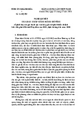
dian year of study recruitment, use of endocrine therapy, use of a Fig. 1.
Scatter plot of unadjusted rates of ipsilateral breast
radiation boost, use of re-excision, ER status, and type of IBTR
tumor recurrence, by median year of study recruitment. Volume 88 Number 3 2014 559
older studies included in this meta-analysis comparing negative
The use of systemic therapy in the treatment of early-stage BC
versus close versus positive margins. Only 26% and 38% of pa-
has changed dramatically over the past 30 years; throughout this
tients included in the entire meta-analysis received chemotherapy
period, consistent evidence has accumulated that successful sys-
and endocrine therapy, respectively, despite a median tumor size
temic therapy improves local control. In the NSABP B06 trial,
of 1.6 cm and a 26% incidence of nodal positivity. Because the
only those women with node-positive disease received chemo-
incidence of local recurrence increases with time, a median
therapy (melphalan and fluorouracil [FU]). Within the cohort that
follow-up of at least 4 years was one of the criteria for inclusion in underwent irradiation, node-positive patients demonstrated
the meta-analysis, and inclusion of studies with a longer follow-up
roughly half the rate of IBTR compared with node-negative pa-
period was believed to be important for an accurate assessment of
tients (5% vs 12%) (2) at 12 years, an advantage attributable to the
the risk of local recurrence. As noted in Fig. 1, the crude incidence
use of chemotherapy. This positive impact of systemic treatment
of IBTR declined over time, and although this was observed for all
on local control has continued with improvements in systemic
margin widths, the decline appeared more pronounced in those
therapy. As illustrated in subsequent analyses of NSABP trials of
with margins <5 mm. As discussed in detail below, the benefits of
systemic therapy, each improvement that led to improved survival
adjuvant systemic therapy in reducing IBTR are well documented
was accompanied by a decline in IBTR. In NSABP B14
(20). The widespread use of systemic therapy today, even for
(tamoxifen vs no tamoxifen for ER-positive, node-negative dis-
patients with small, node-negative BC, increased the confidence of
ease), the rate of IBTR was 11.6% in the control group compared
the MP that wider margins were unlikely to enhance local control
with 5.0% in the tamoxifen group (P<.001) (8); in NSABP B13
in a clinically significant way in the current era. In addition,
(chemotherapy vs not for node-negative disease), IBTR rate was
although the median year of study recruitment of studies included
15.3% in the control and 5.4% in the treated patients (P<.001) (8);
in the meta-analysis was 1990, the median prevalence of IBTR for
in NSABP B19 (methotrexate and FU vs cyclophosphamide,
all studies in the meta-analysis was only 5.3%. Although the ORs
methotrexate, and FU in ER-negative, node-negative patients), the
in Table 4 numerically suggest that 5-mm margins offer an
IBTR rates were 8.2% and 4.8% (P<.03) (25). The 1990s saw the
advantage compared with margins of 1 to 2 mm, these differences
introduction of taxanes into adjuvant and neoadjuvant regimens,
lack statistical significance despite the use of 2 different statistical
and pooled data from NSABP trials B18 (anthracycline-based
tests and robust sample sizes, making it unlikely that the meta-
chemotherapy) and B27 (inclusion of docetaxel) demonstrated
analysis lacks the power to detect clinically meaningful differ-
that women who did not achieve a pathologic complete response
ences in IBTR based on margin width. Furthermore, with the
(pCR) in the breast had an increased hazard ratio (HR) for local-
overall rate of IBTR of 5.3%, the absolute benefit in possible
regional recurrence (HR, 1.55; 95% CI, 1.01-2.59) compared with
decreased IBTR with an OR of 0.77 is on the order of 1% to 2%.
those who did (26). Thus, achieving a pCRdwhich heralds a
More importantly, adjustments for covariates, such as the use of much-reduced risk of distant disease and breast cancer
endocrine therapy and the use of a boost dose of radiation,which
deathdalso results in a significantly reduced risk of IBTR.
are a standard part of practice today, virtually eliminate the
The adjuvant systemic therapy of today is substantially
numeric differences in the ORs (Table 4). Thus, although larger
improved over that of 20 years ago and is likely to continue to
margin widths may have resulted in small reductions in local
improve, with better targeting of specific BC subtypes. For
recurrence in the past, there is no evidence that they are important
women with ER-positive BC undergoing BCT, the 10-year rates
in the setting of current multimodality treatment. It was not
of IBTR in the Early Breast Cancer Trialists’ Group overview
possible to compare rates of IBTR between margins of no ink on
were 18.6% when tamoxifen was not used and 8.7% when
tumor and margins of 1 mm in model 2 (Table 4), because only a
tamoxifen was used (1). The introduction of aromatase inhibitor
small number of studies with these margin definitions were
therapy instead of, or in addition to, tamoxifen in postmenopausal
available for review. The MP considered the long-term results of
women has led to a consistent reduction in the rates of IBTR
the NSABP B06 randomized trial (2), which defined a negative
across essentially all trials, with an average reduction in the HR
margin as no ink on tumor, began accrual in 1976, and reported a
of approximately 0.67 (27), The addition of taxanes to
5% rate of IBTR after 12 years of follow-up in patients receiving
anthracycline-based regimens is also accompanied by a relative
systemic therapy. In addition, the variability in margin assessment
reduction in the rate of IBTR (20). Finally, the addition of tras-
discussed above, the lack of evidence of a significant difference in
tuzumab to cytotoxic regimens for patients with human epidermal
rates of IBTR among margins of 1 mm, 2 mm, and 5 mm, and the
growth factor receptor 2 (HER2)epositive BC leads to a further
benefits of a boost dose of radiation on local control as discussed
reduction in the crude hazard of IBTR, with HRs of 0.47 and 0.66
below led the MP to believe that the totality of evidence did not
in the pooled U.S. trials and European Herceptin Adjuvant
support a distinction between margins of no ink on tumor and
(HERA) trial (28, 29). These data from large randomized clinical margins of 1 mm.
trials establish the principle that systemic therapy advances that
lead to improved survival and decreased risk of distant disease
also contribute to improved local control and suggest that, as
systemic therapy continues to improve, so will its impact on diminishing IBTR.
The panel agreed that the evidence indicates clearly that sys- 3. Systemic therapy
temic therapy, used for the vast majority of patients with BC
The rates of IBTR are reduced with the use of systemic
today, reduces the overall risk of IBTR. It also strengthened the
therapy. In the uncommon circumstance of a patient not
confidence of the MP that 1-mm increments in margin widths are
receiving adjuvant systemic therapy, there is no evidence
unlikely to affect IBTR once a margin of no ink on tumor cells has
suggesting that margins wider than no ink on tumor are
been obtained. Although the evidence base was less robust, the needed.
panel agreed that, in the rare circumstance in which a patient does 560 Moran et al.
International Journal of Radiation Oncology Biology Physics
not receive any form of systemic treatment, there is no evidence to
varies by subtype based on the results of many studies, patients
suggest that obtaining margins wider than no ink on tumor would
with aggressive tumors remain at equally increased risk for local
result in any further reduction of IBTR.
failure irrespective of treatment with mastectomy or BCT, indi-
cating that there is no justification for more widely clear margins
over no ink on tumor for any BC subtype. 4. Biologic subtypes
Margins wider than no ink on tumor are not indicated 5. Radiation therapy delivery based on biologic subtype.
The choice of WBRT delivery technique, fractionation,
and boost dose should not be dependent on margin width.
An improved understanding of biologic subtypes of BC has led
to great improvements in systemic therapy that have, in turn,
decreased IBTR. Several large studies have examined IBTR rates
WBRT options have expanded significantly in the last decade.
with BCT in relation to molecular markers. In 1 of the largest
Delivery techniques such as prone positioning and intensity-
studies, Arvold et al (30) reviewed the cases of 1434 patients who
modulated radiation therapy have been designed to limit
underwent BCT and found that those patients with triple-negative
treatment-related toxicity by decreasing heart/lung volumes and
BC (TNBC) and HER2-positive tumors had a significantly higher
improving homogeneity across the whole-breast radiation field,
risk of IBTR compared with patients with other subtypes. How-
respectively (40-43). In addition, attempts have been made to
ever, the study did not include treatment with adjuvant trastuzu-
decrease the burden of the protracted treatments inherent to
mab, which lowers IBTR for the HER2-positive group. Another
conventionally fractionated WBRT through the use of accelerated,
large study, by Voduc et al (31), of nearly 3,000 patients with a
hypofractionated, whole-breast schemas. Two large randomized
median follow-up of 12 years, also found increased IBTR among
trials have now reported comparable long-term efficacy and
those patients with HER2-enriched and basal tumors. Interest-
toxicity data with these shorter fractionation schedules, estab-
ingly, the investigators found no increased IBTR among TNBCs
lishing it as an acceptable alternative (44, 45). In general, the
with nonbasal tumor markers (31). Mazouni et al (32) reported on
studies evaluating these approaches did not specify particular
1194 patients and found no statistically significant differences in
surgical margin widths, and required only complete microscopic
IBTR on the basis of subtype. They did, however, note that
excision of tumor (40-43, 45). The large United Kingdom Stan-
mastectomy was more commonly performed for HER2-positive
dardization of Breast Radiotherapy (START) trial did mandate a
disease and TNBC than for luminal A and luminal B tumors,
1-mm margin, but comparable long-term results were reported
suggesting that surgeons were less comfortable with BCT for
in the similar Canadian hypofractionation trial that excluded only
more aggressive tumor subtypes, despite a lack of data. Haffty
those with involved margins (45-47). Although neither of these
et al (33), as well as Freedman et al (34), also found no significant
trials was designed to address a possible interaction between
differences in IBTR among patients treated with BCT when
margin width and the specifics of radiation delivery, there is no
comparing TNBC with non-TNBC. A recent study by Gangi et al
evidence to suggest that margin width should dictate patient se-
(35) examined outcome among 1851 consecutive patients treated lection for these therapies.
between 2000 and 2012, during which trastuzumab was routinely
As discussed earlier, a radiation boost to the tumor bed after
used for HER2-positive patients. There was no significant differ-
WBRT has been shown to significantly reduce the risk of IBTR at
ence in IBTR among patients with TNBC compared with other
a cost of increased, although acceptable, rates of late radiation subtypes of tumors.
toxicity (18, 48, 49). In the randomized trials establishing the
Intuitively, it might be thought that wider margins are neces-
benefit of a boost, negative surgical margins were largely defined
sary to control the more aggressive tumor types. However, there is as no ink on tumor.
no reason to believe that HER2-positive disease and TNBC are
Further tailoring of the boost dose has been explored in several
more difficult to resect. Pilewskie et al (36) examined the impact
single-institution series (50-52). In each of these studies, margin
of margin width on local recurrence in 535 patients with TNBC.
width was used as an indicator of potential residual tumor burden,
At 60 months, the incidence of IBTR did not differ significantly
and boost doses were increased with decreasing margin width.
between patients with margins 2 mm and those with margins >2
The MP believed that interpretation of these and other retro-
mm (7.3% vs 5.1%). Alternatively, local failure occurs as a marker
spective data evaluating both radiation dose and surgical margins
of aggressive biology, as is seen after mastectomy. Three retro-
was complicated by the heterogeneity of total radiation doses and
spective studies have examined the incidence of local failure in
techniques and by a lack of control cohorts with comparable
TNBC after BCT or mastectomy, and have found no difference
margin widths and uniform doses. Therefore, the panel concluded
based on surgical procedure, suggesting that these local re-
that there was no clear reduction in IBTR as a result of escalating
currences are more likely a result of aggressive biology, not re-
the radiation dose when margin widths were smaller. In one report,
sidual tumor at the surgical site, which could be improved with
an increased rate of IBTR was noted in patients with close or
wider lumpectomy margins (29, 37-39). This theory is supported
positive margins despite the dose-escalation strategy (51). The
by the approximately 40% decline in IBTR seen in patients with
other studies simply noted the lack of a clear relationship between
HER2-positive tumors receiving adjuvant systemic trastuzumab
local control and margin width or radiation dose (50, 52).
and other HER2-targeted agents (29). In summary, the MP
In summary, margin width should not be used to determine the
concluded that, although there is evidence that the risk of IBTR
delivery technique or fractionation for WBRT or vice versa. Volume 88 Number 3 2014 561
Furthermore, in patients with negative margins (no ink on tumor),
in older women. In the Early Breast Cancer Trialists’ Collaborative
the use and dose of a tumor bed boost should be based on a priori
Group meta-analysis of breast-conserving surgery with and without
estimation of local failure risk and should not be determined, in
radiation therapy, the rate of any first recurrence by age was 5.9% per
year for age <40 years, 2.7% per year for age 40 to 49 years, and 1%
to 1.9% per year for 50 years in the node-negative subgroup (1).
Corresponding rates in the node-positive subgroup were 8.3% per
year for age <40 years, 6.5% per year for age 40 to 49 years, and 4.8%
6. Invasive lobular carcinoma and lobular
to 6.5% per year for age 50 years, respectively. An increased risk for carcinoma in situ
BC mortality was also seen in the subgroup of women aged <40
years. Other studies have confirmed a higher risk of distant recurrence
Wider negative margins than no ink on tumor are not
as well as IBTR in young women (61, 62).
indicated for invasive lobular carcinoma (ILC). Classic
Young patient age is not associated with an improved outcome
lobular carcinoma in situ (LCIS) at the margin is not an
with mastectomy. The risk for locoregional recurrence after
indication for re-excision. The significance of pleomor-
mastectomy without radiation is also significantly higher in young
phic LCIS at the margin is uncertain.
women compared with their older counterparts (63), and the
increased risk of both recurrence and BC death is not improved
isolation, by the width of the surgical margin.
with mastectomy compared with BCT (62, 64). The increased
IBTR rates in young women likely result from the greater fre-
ILCs comprise 5% to 15% of all BCs. Several large retro-
quency of adverse biologic and pathologic features in this group
spective studies have demonstrated that when negative margins
compared with older women. Young women have more aggressive
were obtained, the risk of IBTR was not significantly different
tumor characteristics, such as high histologic grade, lymphovas-
between ILC and invasive ductal carcinoma (53-55). Wider mar-
cular invasion, hormone receptorenegative BC, BRCA1, and
gins do not yield lower IBTR rates. In a retrospective study of 382
BRCA2 mutationeassociated cancers, and BCs associated with
patients comparing margins >1 cm with smaller margins, no
adverse gene expression profiles (65, 66) compared with their
differences in local recurrence rates were observed (56). In
older counterparts. In 1 study, very young patients with tumors
addition, most classical ILCs have a luminal A phenotype and are
classified as luminal B, HER2, and triple-negative subtypes were
ER positive, so the benefits of endocrine therapy on local control,
at increased risk for IBTR when compared with older patients, but
as discussed previously, will be seen in this population. Thus, the
no significant effect of age was seen in the subgroup with the most
MP concluded that the general recommendations regarding
favorable luminal A subtype (66). Young age may be a less
margin width should not be altered for invasive lobular histology.
important factor for IBTR when controlling for adverse gene-
In contrast to clear evidence demonstrating that DCIS at the
expression profile (30, 67) or may not be important at all in pre-
margin increases IBTR, the presence of LCIS at the margin does
dicting recurrence and survival in an era of modern systemic
not affect IBTR. In a retrospective study, the 10-year cumulative
therapy and anti-HER2edirected therapy, as suggested in 1 recent
incidence rate of IBTR in patients with BC was not significantly
study of young women with HER2-overexpressing cancers (68).
different in patients with or without LCIS unless tamoxifen was
There was no evidence in the margins meta-analysis that, once a
withheld (57). In other large studies, the presence of LCIS within
negative margin has been achieved, young patients benefit from a
the specimen or at the resection margin did not appear to affect the
greater negative margin width than no ink on tumor. In 18 studies in
risk of local recurrence (58, 59). There is concern that the pleo-
the meta-analysis, the adjusted OR for IBTR with age as a covariate
morphic variant of LCIS, which has some features more akin to
did not differ significantly when negative margin widths were defined
high-grade DCIS than to classical LCIS, may carry an increased
as 1 mm, 2 mm, or 5 mm (P for association, .86; P for trend, .58). This
risk of recurrence when at the margin. Given the limitation of only
is consistent with the finding that mastectomy, which theoretically
small retrospective studies with a very limited number of events
should provide the largest margin width that can be obtained, is also
available to address this question (60), the MP did not believe that
associated with an increased risk of local recurrence in younger
a recommendation regarding pleomorphic LCIS at the margin
compared with older women. In addition, there are data demon- could be made at this time.
strating equivalent risks for recurrence and BC death in young
women irrespective of treatment with BCT or mastectomy (62, 64).
Thus, the MP concluded that although the adverse pathologic
and biologic factors associated with young age are mitigated to
some extent by excision to negative margins, use of systemic
therapies, use of a radiation boost, and possible exclusion of 7. Young age
young BRCA mutation carriers from a BCT approach, there is no
Young age (40 years) is associated with both increased
IBTR after BCT as well as increased local relapse on the
chest wall after mastectomy, and is also more frequently
associated with adverse biologic and pathologic features. 8. Lobular carcinoma in situ
There is no evidence that increased margin width nullifies
the increased risk of IBTR in young patients.
A lobular carcinoma in situ (EIC) identifies patients who
may have a large residual DCIS burden after lumpec-
tomy. There is no evidence of an association between
Young patient age, usually defined as <40 years, has been asso-
increased risk of IBTR and EIC when margins are
ciated with an increased risk of IBTR after BCT compared with that negative. 562 Moran et al.
International Journal of Radiation Oncology Biology Physics
evidence supporting obtaining wider negative margins beyond no
5. McCahill LE, Single RM, Aiello Bowles EJ, et al. Variability in
ink on tumor solely on the basis of young patient age.
reexcision following breast conservation surgery. JAMA 2012;307: 467-475.
EIC is a pathologic description of invasive ductal carcinoma
6. Morrow M, Jagsi R, Alderman AK, et al. Surgeon recommendations
and receipt of mastectomy for treatment of breast cancer. JAMA
that has a prominent intraductal component within the tumor and 2009;302:1551-1556.
adjacent normal tissue. The basis of the definition of EIC was the
7. King TA, Sakr R, Patil S, et al. Clinical management factors
observation in the 1970s at the Harvard Joint Center for Radiation
contribute to the decision for contralateral prophylactic mastectomy.
Therapy, at a time when margins of resection were not routinely
J Clin Oncol 2011;29:2158-2164.
assessed, that a high rate of IBTR was observed in patients un-
8. Anderson S, Wapnir I, Dignam J, et al. Prognosis after ipsilateral
dergoing BCT when a prominent DCIS burden was noted within
breast tumor recurrence and locoregional recurrences in patients
the confines of the invasive cancer (approximately 25%) and
treated by breast-conserving therapy in five National Surgical
within breast tissue beyond the edges of the invasive cancer (69).
Adjuvant Breast and Bowel Project protocols of node-negative breast
These EIC-positive cancers often recurred within or at the edge of
cancer. J Clin Oncol 2009;27:2466-2473.
the boost volume and were more commonly seen in young patients
9. Graham R, Mancher M, Wolman DM, et al, editors. Clinical Practice
Guidelines We Can Trust. Washington, DC: National Academies
(35 years of age). Furthermore, IBTR was more common in Press; 2011.
young EIC-positive patients than in older EIC-positive patients.
10. Sardanelli F, Podo F, Santoro F, et al. Multicenter surveillance of
In subsequent years, when margins of resection were inked, and
women at high genetic breast cancer risk using mammography, ul-
re-excisions were performed for positive or close margins, patients
trasonography, and contrast-enhanced magnetic resonance imaging
with EIC-positive cancers (but not EIC-negative cancers) were
(the High Breast Cancer Risk Italian 1 Study): Final results. Invest
frequently found to have considerable residual DCIS in the re- Radiol 2011;46:94-105.
excision specimens (70). Pathologic examination of a cohort of
11. Moher D, Liberati A, Tetzlaff J, et al. Preferred reporting items for
mastectomy specimens revealed that 33% of EIC-positive cancers
systematic reviews and meta-snalyses: The PRISMA statement. J
had prominent DCIS (6 low-power fields of DCIS) at 2 cm from
Clin Epidemiol 2009;62:1006-1012.
the edge of the index cancer compared with only 2% of EIC-negative
12. Houssami N, Macaskill P, Marinovich ML, et al. Meta-analysis of the
impact of surgical margins on local recurrence in women with early-
cancers (71). In aggregate, these studies indicated that an EIC de-
stage invasive breast cancer treated with breast-conserving therapy.
notes a cancer that may have extensive multifocal DCIS involve-
Eur J Cancer 2010;46:3219-3232.
ment and an increased rate of IBTR if not adequately resected.
13. Houssami N, Macaskill P, Marinovich ML, et al. The association of
Later, additional studies revealed that patients with EIC-
surgical margins and local recurrence in women with early stage
positive tumors did not have an increase in IBTR unless tumor
invasive breast cancer treated with breast conserving therapy: A
cells were present at the inked margin (72). In a cohort of EIC-
metaanalysis. Ann Surg Oncol. In press.
positive patients, IBTR was 0% at 5 years when there were no
14. Bellon JR, Come SE, Gelman RS, et al. Sequencing of chemotherapy
tumor cells at the inked margin or when the margin was defined as
and radiation therapy in early-stage breast cancer: Updated results of
close, but it was 50% when there was more than focal positivity
a prospective randomized trial. J Clin Oncol 2005;23:1934-1940.
(72). On the basis of this information, the MP did not believe that
15. Voogd AC, Nielsen M, Peterse JL, et al. Differences in risk factors
for local and distant recurrence after breast-conserving therapy or
the available evidence supports the routine use of margins wider
mastectomy for stage I and II breast cancer: Pooled results of two
than no ink on tumor. However, in view of the potential for sub-
large European randomized trials. J Clin Oncol 2001;19:1688-1697.
stantial residual DCIS in EIC-positive patients, consideration
16. Wazer DE, Schmidt-Ullrich RK, Ruthazer R, et al. Factors deter-
should be given to obtaining postoperative mammographic im-
mining outcome for breast-conserving irradiation with margin-
aging to assist in identifying residual tumor bed calcifications
directed dose escalation to the tumor bed. Int J Radiat Oncol Biol
warranting re-excision. In addition, when an EIC is present, young Phys 1998;40:851-858.
age and multiple close margins are associated with an increased
17. Mansfield CM, Komarnicky LT, Schwartz GF, et al. Ten-year results
risk of IBTR and can be used to select patients who might benefit
in 1070 patients with stages I and II breast cancer treated by con-
from re-excision (69, 72). Postexcision mammography is a useful
servative surgery and radiation therapy. Cancer 1995;75:2328-2336.
adjunct to margin status to assess the completeness of excision of
18. Bartelink H, Horiot JC, Poortmans PM, et al. Impact of a higher
radiation dose on local control and survival in breast-conserving
lesions with calcifications even when an EIC is not present.
therapy of early breast cancer: 10-Year results of the randomized
boost versus no boost EORTC 22881-10882 trial. J Clin Oncol 2007; References 25:3259-3265.
19. Jones HA, Antonini N, Hart AA, et al. Impact of pathological
characteristics on local relapse after breast-conserving therapy: A
1. Darby S, McGale P, Correa C, et al. Effect of radiotherapy after
subgroup analysis of the EORTC boost versus no boost trial. J Clin
breast-conserving surgery on 10-year recurrence and 15-year breast Oncol 2009;27:4939-4947.
cancer death: Meta-analysis of individual patient data for 10,801
20. Mannino M, Yarnold JR. Local relapse rates are falling after breast
women in 17 randomised trials. Lancet 2011;378:1707-1716.
conserving surgery and systemic therapy for early breast cancer: Can
2. Fisher B, Anderson S, Bryant J, et al. Twenty-year follow-up of a
radiotherapy ever be safely withheld? Radiother Oncol 2009;90:14-22.
randomized trial comparing total mastectomy, lumpectomy, and
21. Holland R, Veling SH, Mravunac M, et al. Histologic multifocality of
lumpectomy plus irradiation for the treatment of invasive breast
Tis, T1-2 breast carcinomas: Implications for clinical trials of breast-
cancer. N Engl J Med 2002;347:1233-1241.
conserving surgery. Cancer 1985;56:979-990.
3. Azu M, Abrahamse P, Katz SJ, et al. What is an adequate margin for
22. Graham RA, Homer MJ, Katz J, et al. The pancake phenomenon
breast-conserving surgery? Surgeon attitudes and correlates. Ann
contributes to the inaccuracy of margin assessment in patients with Surg Oncol 2010;17:558-563.
breast cancer. Am J Surg 2002;184:89-93.
4. Taghian A, Mohiuddin M, Jagsi R, et al. Current perceptions
23. Guidi AJ, Connolly JL, Harris JR, et al. The relationship between
regarding surgical margin status after breast-conserving therapy:
shaved margin and inked margin status in breast excision specimens.
Results of a survey. Ann Surg 2005;241:629-639. Cancer 1997;79:1568-1573. Volume 88 Number 3 2014 563
24. Carter D. Margins of “lumpectomy” for breast cancer. Hum Pathol
46. Bentzen SM, Agrawal RK, Aird EG, et al. The UK Standardisation of 1986;17:330-332.
Breast Radiotherapy (START) Trial A of radiotherapy hypofractio-
25. Fisher B, Jeong JH, Anderson S, et al. Treatment of axillary lymph
nation for treatment of early breast cancer: A randomised trial.
node-negative, estrogen receptor-negative breast cancer: Updated Lancet Oncol 2008;9:331-341.
findings from National Surgical Adjuvant Breast and Bowel Project
47. Bentzen SM, Agrawal RK, Aird EG, et al. The UK Standardisation of
clinical trials. J Natl Cancer Inst 2004;96:1823-1831.
Breast Radiotherapy (START) Trial B of radiotherapy hypofractio-
26. Mamounas EP, Anderson SJ, Dignam JJ, et al. Predictors of
nation for treatment of early breast cancer: A randomised trial.
locoregional recurrence after neoadjuvant chemotherapy: Results Lancet 2008;371:1098-1107.
from combined analysis of National Surgical Adjuvant Breast and
48. Polga´r C, Fodor J, Orosz Z, et al. Electron and high-dose-rate
Bowel Project B-18 and B-27. J Clin Oncol 2012;30:3960-3966.
brachytherapy boost in the conservative treatment of stage I-II
27. Hind D, Ward S, De Nigris E, et al. Hormonal therapies for early
breast cancer: First results of the randomized Budapest Boost Trial.
breast cancer: Systematic review and economic evaluation. Health
Strahlenther Onkol 2002;178:615-623.
Technol Assess 2007;11:1-134. iii-iv, ix-xi.
49. Romestaing P, Lehingue Y, Carrie C, et al. Role of a 10-Gy boost in
28. Smith I, Procter M, Gelber RD, et al. 2-Year follow-up of trastuzu-
the conservative treatment of early breast cancer: Results of a ran-
mab after adjuvant chemotherapy in HER2-positive breast cancer: A
domized clinical trial in Lyon, France. J Clin Oncol 1997;15:963-
randomised controlled trial. Lancet 2007;369:29-36. 968.
29. Romond EH, Perez EA, Bryant J, et al. Trastuzumab plus adjuvant
50. Livi L, Meattini I, Franceschini D, et al. Radiotherapy boost dose-
chemotherapy for operable HER2-positive breast cancer. N Engl J
escalation for invasive breast cancer after breast-conserving sur- Med 2005;353:1673-1684.
gery: 2093 Patients treated with a prospective margin-directed policy.
30. Arvold ND, Taghian AG, Niemierko A, et al. Age, breast cancer
Radiother Oncol 2013;108:273-278.
subtype approximation, and local recurrence after breast-conserving
51. Neuschatz AC, DiPetrillo T, Safaii H, et al. Long-term follow-up of a
therapy. J Clin Oncol 2011;29:3885-3891.
prospective policy of margin-directed radiation dose escalation in
31. Voduc KD, Cheang MC, Tyldesley S, et al. Breast cancer subtypes and
breast-conserving therapy. Cancer 2003;97:30-39.
the risk of local and regional relapse. J Clin Oncol 2010;28:1684-1691.
52. Murphy C, Anderson PR, Li T, et al. Impact of the radiation boost on
32. Mazouni C, Rimareix F, Mathieu MC, et al. Outcome in breast
outcomes after breast-conserving surgery and radiation. Int J Radiat
molecular subtypes according to nodal status and surgical pro- Oncol Biol Phys 2011;81:69-76.
cedures. Am J Surg 2013;205:662-667.
33. Haffty BG, Yang Q, Reiss M, et al. Locoregional relapse and distant
53. Biglia N, Maggiorotto F, Liberale V, et al. Clinical-pathologic fea-
metastasis in conservatively managed triple-negative early-stage
tures, long term-outcome and surgical treatment in a large series of
breast cancer. J Clin Oncol 2006;24:5652-5657.
patients with invasive lobular carcinoma (ILC) and invasive ductal
34. Freedman GM, Anderson PR, Li T, et al. Locoregional recurrence of
carcinoma (IDC). Eur J Surg Oncol 2013;39:455-460.
triple-negative breast cancer after breast-conserving surgery and ra-
54. Chung MA, Cole B, Wanebo HJ, et al. Optimal surgical treatment of
diation. Cancer 2009;115:946-951.
invasive lobular carcinoma of the breast. Ann Surg Oncol 1997;4:
35. Gangi A, Chung A, Mirocha J, Liou DZ, Leong T, Giuliano AE. 545-550.
Breast-Conserving Therapy for Triple-Negative Breast Cancer.
55. Winchester DJ, Chang HR, Graves TA, et al. A comparative analysis
JAMA surg 2014 Jan 1 [Epub ahead of print]. http://dx.doi.org/10.
of lobular and ductal carcinoma of the breast: Presentation, treat- 1001/jamasurg.2013.3037.
ment, and outcomes. J Am Coll Surg 1998;186:416-422.
36. Pilewskie ML. Effect of margin width on local recurrence (LR) in
56. Galimberti V, Maisonneuve P, Rotmensz N, et al. Influence of margin
triple-negative breast cancer treated with breast-conserving surgery
status on outcomes in lobular carcinoma: Experience of the European
(BCT). J Clin Oncol 2013;31(15S):54.
Institute of Oncology. Ann Surg 2011;253:580-584.
37. Abdulkarim BS, Cuartero J, Hanson J, et al. Increased risk of
57. Sasson AR, Fowble B, Hanlon AL, et al. Lobular carcinoma in situ
locoregional recurrence for women with T1-2N0 triple-negative
increases the risk of local recurrence in selected patients with stages I
breast cancer treated with modified radical mastectomy without
and II breast carcinoma treated with conservative surgery and radi-
adjuvant radiation therapy compared with breast-conserving therapy.
ation. Cancer 2001;91:1862-1869.
J Clin Oncol 2011;29:2852-2858.
58. Ciocca RM, Li T, Freedman GM, et al. Presence of lobular carci-
38. Ho AY, Gupta G, King TA, et al. Favorable prognosis in patients with
noma in situ does not increase local recurrence in patients treated
T1a/T1bN0 triple-negative breast cancers treated with multimodality
with breast-conserving therapy. Ann Surg Oncol 2008;15:2263-2271.
therapy. Cancer 2012;118:4944-4952.
59. Moran M, Haffty BG. Lobular carcinoma in situ as a component of
39. Adkins FC, Gonzalez-Angulo AM, Lei X, et al. Triple-negative
breast cancer: The long-term outcome in patients treated with
breast cancer is not a contraindication for breast conservation. Ann
breast-conservation therapy. Int J Radiat Oncol Biol Phys 1998;40: Surg Oncol 2011;18:3164-3173. 353-358.
40. Formenti SC, Gidea-Addeo D, Goldberg JD, et al. Phase I-II trial of
60. Downs-Kelly E, Bell D, Perkins GH, et al. Clinical implications of
prone accelerated intensity modulated radiation therapy to the breast
margin involvement by pleomorphic lobular carcinoma in situ. Arch
to optimally spare normal tissue. J Clin Oncol 2007;25:2236-2242.
Pathol Lab Med 2011;135:737-743.
41. Donovan E, Bleakley N, Denholm E, et al. Randomised trial of
61. Fisher ER, Anderson S, Tan-Chiu E, et al. Fifteen-year prognostic
standard 2D radiotherapy (RT) versus intensity modulated radio-
discriminants for invasive breast carcinoma: National Surgical
therapy (IMRT) in patients prescribed breast radiotherapy. Radiother
Adjuvant Breast and Bowel Project Protocol-06. Cancer 2001; Oncol 2007;82:254-264. 91(Suppl.):1679-1687.
42. Formenti SC, DeWyngaert JK, Jozsef G, et al. Prone vs supine
62. de Bock GH, van der Hage JA, Putter H, et al. Isolated loco-regional
positioning for breast cancer radiotherapy. JAMA 2012;308:861-863.
recurrence of breast cancer is more common in young patients and
43. Pignol JP, Olivotto I, Rakovitch E, et al. A multicenter randomized
following breast conserving therapy: Long-term results of European
trial of breast intensity-modulated radiation therapy to reduce acute
Organisation for Research and Treatment of Cancer studies. Eur J
radiation dermatitis. J Clin Oncol 2008;26:2085-2092. Cancer 2006;42:351-356.
44. Haviland J, Agrawal R, Aird E, et al. The UK START (Stand-
63. Taghian A, Jeong J, Mamounas E, et al. Patterns of locoregional
ardisation of Breast Radiotherapy) trials: 10-Year follow-up results
failure in patients with operable breast cancer treated by mastectomy
[abstract S4-1]. Cancer Res 2012;72(Suppl.):24.
and adjuvant chemotherapy with or without tamoxifen and without
45. Whelan TJ, Pignol JP, Levine MN, et al. Long-term results of
radiotherapy: Results from five national Surgical Adjuvant Breast
hypofractionated radiation therapy for breast cancer. N Engl J Med
and Bowel Project randomized clinical trials. J Clin Oncol 2004;22: 2010;362:513-520. 4247-4254. 564 Moran et al.
International Journal of Radiation Oncology Biology Physics
64. Kroman N, Holtveg H, Wohlfahrt J, et al. Effect of breast-conserving
factor receptor 2epositive breast cancer: Results from a herceptin
therapy versus radical mastectomy on prognosis for young women
adjuvant trial. J Clin Oncol 2013;31:2692-2698.
with breast carcinoma. Cancer 2004;100:688-693.
69. Schnitt SJ, Connolly JL, Harris JR, et al. Pathologic predictors of
65. Anders CK, Hsu DS, Broadwater G, et al. Young age at diagnosis
early local recurrence in stage I and II breast cancer treated by pri-
correlates with worse prognosis and defines a subset of breast cancers
mary radiation therapy. Cancer 1984;53:1049-1057.
with shared patterns of gene expression. J Clin Oncol 2008;26:3324-
70. Schnitt SJ, Connolly JL, Khettry U, et al. Pathologic findings on re- 3330.
excision of the primary site in breast cancer patients considered for
66. Cancello G, Maisonneuve P, Rotmensz N, et al. Prognosis and
treatment by primary radiation therapy. Cancer 1987;59:675-681.
adjuvant treatment effects in selected breast cancer subtypes of very
71. Holland R, Connolly JL, Gelman R, et al. The presence of an
young women (<35 years) with operable breast cancer. Ann Oncol
extensive intraductal component following a limited excision corre- 2010;21:1974-1981.
lates with prominent residual disease in the remainder of the breast. J
67. Demirci S, Broadwater G, Marks LB, et al. Breast conservation Clin Oncol 1990;8:113-118.
therapy: The influence of molecular subtype and margins. Int J
72. Schnitt SJ, Abner A, Gelman R, et al. The relationship between
Radiat Oncol Biol Phys 2012;83:814-820.
microscopic margins of resection and the risk of local recurrence in
68. Partridge AH, Gelber S, Piccart-Gebhart MJ, et al. Effect of age on
patients with breast cancer treated with breast-conserving surgery
breast cancer outcomes in women with human epidermal growth
and radiation therapy. Cancer 1994;74:1746-1751.
Document Outline
- Society of Surgical Oncology–American Society for Radiation Oncology Consensus Guideline on Margins for Breast-Conserving S ...
- Introduction
- Methods and Materials
- Literature review and meta-analysis
- Inclusion/exclusion criteria
- Study quality and limitations of the literature
- Management of conflicts of interest for the MP
- Results
- Guideline recommendations
- 1. Positive margins
- 2. Negative margin widths
- 3. Systemic therapy
- 5. Radiation therapy delivery
- 8. Lobular carcinoma in situ
- References
Tài liệu liên quan:
-

Ung dung game hoa trong cac chien dich MKT
37 19 -

Bao cao Chi so TMDT Viet Nam 2025
37 19 -

Thông tư quy định về việc phân quyền, phân cấp và phân định thẩm quyền quản lý nhà nước về giáo dục cho chính quyền địa phương
42 21 -
Nghị quyết về phát huy các giá trị di sản văn hóa gắn với phát triên du lịch bền vững tỉnh Khánh Hòa đến năm 2025, định hướng đến năm 2030
41 21 -

Quyết định phê duyệt Chiến lược phát triển du lịch Việt Nam đến năm 2030
26 13