Zhang 2020 Môn Xác suất thống kê y học | Trường Đại học Huế
The members of the organic anion transporter (OAT) family are mainly expressed in kidney, liver, placenta, intes-tine, and brain. These transporters play important roles in the disposition of clinical drugs, pesticides, signalingmolecules, heavy metal conjugates, components of phytomedicines, and toxins, and therefore critical for main-taining systemic homeostasis. Tài liệu gồm 14 trang, giúp các bạn tham khảo, ôn tập và đạt kết quả cao. Mình bạn đọc đón xem!
Môn: Xác suất thống kê y học (ĐHH) 10 tài liệu
Trường: Đại học Huế 411 tài liệu
Tác giả:


Preview text:
Pharmacology & Therapeutics xxx (xxxx) xxx
Contents lists available at ScienceDirect
j o u r na l h om ep a ge : ww w. e ls e v i e r. c om / loc at e/ p h arm t he ra
Regulation of organic anion transporters: Role in physiology,
pathophysiology, and drug elimination
Jinghui Zhang, Haoxun Wang, Yunzhou Fan, Zhou Yu, Guofeng You ⁎
Department of Pharmaceutics, Rutgers, the State University of New Jersey, Piscataway, NJ, USA A R T I C L E I N f О A B S T R A C T Available online xxxx
The members of the organic anion transporter (OAT) family are mainly expressed in kidney, liver, placenta, intes-
tine, and brain. These transporters play important roles in the disposition of clinical drugs, pesticides, signaling Keywords:
molecules, heavy metal conjugates, components of phytomedicines, and toxins, and therefore critical for main- Drug transporter
taining systemic homeostasis. Alterations in the expression and function of OATs contribute to the intra- and Organic anion transporter
inter-individual variability of the therapeutic efficacy and the toxicity of many drugs, and to many pathophysio- Drug disposition
logical conditions. Consequently, the activity of these transporters must be highly regulated to carry out their
Post-translational modification
normal functions. This review will present an update on the recent advance in understanding the cellular and Regulation
molecular mechanisms underlying the regulation of renal OATs, emphasizing on the post-translational modifica-
tion (PTM), the crosstalk among these PTMs, and the remote sensing and signaling network of OATs. Such knowl-
edge will provide significant insights into the roles of these transporters in health and disease.
© 2020 Elsevier Inc. All rights reserved. Contents 1.
Introduction ........................................................................................................................................................................................... 0 2.
OAT expression, structure, and function ........................................................................................................................................... 0 3.
OATs and drug-drug interaction (DDI) ............................................................................................................................................... 0 4.
OATs in kidney injury and diseases ................................................................................................................................................... 0 5.
Genetic polymorphisms of OATs and clinical impact ...................................................................................................................... 0 6.
Roles of OATs in the handling of endogenous substances and their metabolites ....................................................................... 0 7.
Regulations of OATs .............................................................................................................................................................................. 0 8.
Conclusion .............................................................................................................................................................................................. 0
Acknowledgements ........................................................................................................................................................................................ 0
Reference ......................................................................................................................................................................................................... 0 1. Introduction
multiple tissues, such as kidney, liver, brain, placenta, retina, and olfac-
tory mucosa (He et al., 2014; S. K. Nigam et al., 2015). They are the key
Organic anion transporters (OATs), a subfamily of the solute carrier
players for the translocation of various substances into and out of cells,
22 (SLC22) transporters, are localized on the physiological barriers of
such as signaling molecules, toxins, and a diverse array of important
clinical therapeutics, including antivirals, anti-cancer drugs, antibiotics,
anti-hypertensives, and anti-inflammatories. (Ahn & Nigam, 2009; Cha
Abbreviations: OAT, organic anion transporter; TMD, transmembrane domain; DDI,
et al., 2000; Dantzler & Wright, 2003; He et al., 2014; Pritchard, 1990;
drug–drug interaction; Nedd4-1/Nedd4-2, neural precursor cell expressed developmen-
Srimaroeng, Perry, & Pritchard, 2008; Taki, Nakamura, Miglinas, tally down-regulated 4-1/4-2; PTM, post-translational modification; DUB,
deubiquitinating enzyme; Sgk, serum- and glucocorticoid-inducible kinase; PKA, protein
Enomoto, & Niwa, 2006; Terada & Inui, 2007; Vallon et al., 2008;
Kinase A; PKB, protein Kinase B; PKC, protein Kinase C; USP8, ubiquitin-specific protease
VanWert, Gionfriddo, & Sweet, 2010; You, 2002). Therefore, OATs are
8; SENP, SUMO1/sentrin specific peptidase; IGF-1, Insulin-like growth factor 1.
not only critical for physiological and pathological processes in the
* Corresponding author at: Department of Pharmaceutics, Rutgers, The State University
body, but also critical in absorption, distribution, metabolism, and
of New Jersey, 160 Frelinghuysen Road, Piscataway, NJ 08854, USA.
E-mail address: gyou@pharmacy.rutgers.edu (G. You).
elimination (ADME) of clinical therapeutics, thus affecting the https://doi.org/10.1 016/j.pharmthera.20 20.107647 0163- 7258/© 2020 Elsevier Inc. All rights reserved.
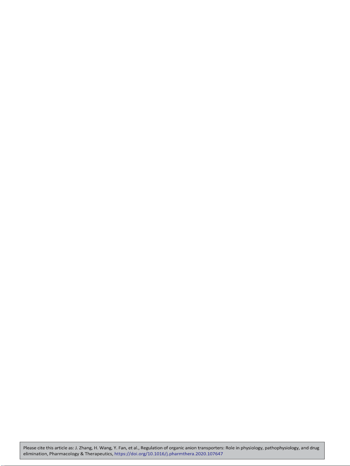
Fig. 1. Major drug transporters expressed in human renal proximal tubule cells. MRP: multidrug resistance-associated protein, OCT: organic cation transporter, OAT: organic anion
transporter, OATP: organic anion-transporting peptide, MATE: multidrug and toxin extrusion protein, PEPT: peptide transporter, BCRP: breast cancer resistance protein, MDR:
multidrug resistance mutation, URAT: urate transporter.
pharmacokinetics and pharmacodynamics of the drug profile. Among
2012; Koepsell, 2013; S. K. Nigam et al., 2015; L. Wang & Sweet,
the tissues involved in the ADME of clinical therapeutics, the kidney is
2013b; D. Xu, Wang, & You, 2016b).
one of the vital organs responsible for drug elimination after its admin-
Kidney is responsible for eliminating substances from the blood and
istration. Renal drug transporters are in charge of the transfer of the
reabsorbing certain compounds back into the circulation. In this way it
drugs between blood and proximal tubule lumen. Among various
keeps essential nutrients in the circulation while removing harmful me-
renal drug transporters, OATs, mainly interacting with organic anionic
tabolites and therapeutic agents from the body (L. Wang & Sweet,
molecules, are expressed at both the basolateral membrane and apical
2013b). There are three major events in this process: glomerulus filtra-
membrane of the proximal tubule cells and are responsible for the ex-
tion, tubule secretion, and reabsorption. Unlike the passive filtration
cretion of numerous endogenous and exogenous substances. Because
processes occurring in kidney glomeruli, much of the active exchange
of the importance of OATs in disposition of many important clinical
of compounds happen in the kidney proximal tubule, where a number
drugs and in various physio-pathological processes, numerous efforts
of important transporters are expressed (Fig. 1). Proximal tubule cells
have been made to uncover molecular and cellular mechanisms that
polarize into apical membrane, which faces the urine, and basolateral
contribute to the regulation of OATs. In this review, we discussed the re-
membrane, which faces the blood vessel. The OAT members are
cent advance in understanding the regulation of OATs, highlighting the
expressed in renal proximal tubule, both on the basolateral and on the
regulation at the level of post-translational modification and the regula-
apical membrane. They stand as important mediators in the active pro-
tory network of the remote sensing and signaling.
cess of renal elimination and reabsorption (Emami Riedmaier et al.,
2012; Motohashi et al., 2013).
2. OAT expression, structure, and function
In human, OAT1, OAT2, OAT3, OAT4, OAT10, and URAT1 have been
detected in kidney samples. It is worth to mention that in rodents cer-
The OAT family consists of a group of transmembrane proteins with
tain Oats have different expression profiles from those in human.
around 540-560 amino acids. The OATs have been identified in the epi-
These comparisons could help to understand the differences of OAT
thelia barriers such as kidney, liver, brain, placenta, and intestine. These
functions and regulations between rodent models and clinical studies.
OATs play significant roles in modulating the movement of organic
(S. K. Nigam et al., 2015; L. Wang & Sweet, 2013b). OAT1, OAT2, and
anion molecules across cell membranes. One of the characteristics of
OAT3 have been located on the basolateral membrane of the proximal
OATs is their wide range of substrate recognition including both physi-
tubule cells in human kidney, which is the blood side of these cells
ological/endogenous substrates and their metabolites and xenobiotic
(Fig. 1). These three OATs facilitate the transfer of organic anions from
molecules such as environmental toxins and therapeutic drugs, which
blood into the proximal tubule cells driven by a tertiary transporting
makes them important players in body homeostasis and pharmacolog-
mechanism. Across basolateral membrane, the cells utilize the sodium
ical responses (VanWert et al., 2010). Understanding the relationships
gradient generated by Na+-K+-ATPase to indirectly drive the influx of
between the molecular features of OATs and their functions provided
organic anions into the proximal tubule cells. OAT4 and URAT1 (another
significant insights into their influences on clinical drug elimination, ef-
member of OAT family) are expressed on the apical membrane or the
ficacy, and toxicity. (Emami Riedmaier, Nies, Schaeffeler, & Schwab,
urine side of the proximal tubule cells (Fig. 1). OAT4 mediates
reabsorption of organic anions from urine back into the tubule cells.
enhanced toxicity and increased risk of developing rhabdomyolysis
OAT10, with higher expression in kidney proximal tubule and small in-
(Feng et al., 2016). In addition to approved drugs, indoxyl sulfate, a ure-
testine, has been proved to transport nicotine and uric acid. URAT1 is
mic toxin, was confirmed as a substrate of OAT1, OAT3, and OAT4 in cul-
mainly responsible for the reabsorption of urate via monocarboxylates
tured cells (Hsueh et al., 2016). In a rat study, the blood concentration of
exchange. (S. K. Nigam et al., 2015; L. Wang & Sweet, 2013b).
indoxyl sulfate increased with lower renal clearance when adminis-
Through computer modeling and site-directed mutagenesis studies,
trated with quinapril, whose metabolite quinaprilat was also a substrate
it has been revealed that OAT family shares a similar structure feature
of Oat3 (Fujita et al., 2012; Yuan et al., 2009). To connect in vitro studies
consisting of intracellular N- and C-termini, 12 α-helical transmem-
with in vivo research, Mathialagan, et al. applied transporter data ob-
brane domains (TMDs), a large extracellular loop between TMD1/2,
tained from OAT-expressing human cells to quantitatively predict
and a central intracellular loop between TMD6/7 (Anzai, Kanai, &
in vivo renal elimination and total renal clearance. Their results con-
Endou, 2006; F. Zhou & You, 2007; Zhu et al., 2015). Three highly con-
cluded that renal transport mediated by OAT3 played a predominant
served regions are mainly responsible for protein functions: the large
role in renal elimination for a majority of drugs they tested
extracellular domain between TMD1/2, the central intracellular loop be- (Mathialagan et al., 2017).
tween TMD6/7, and TMD9/10. These three regions work together to en-
Recognized as a powerful organic anion transport inhibitor, proben-
sure substrate specificity and proper transport activity of OATs
ecid was used with clinical therapies to prolong half-life of drugs there-
(Koepsell, 2013; Zhu et al., 2015). Furthermore, the large extracellular
fore enhance therapeutic effects (L. Wang & Sweet, 2013b). In healthy
domain between TMD1/2 contains multiple glycosylation sites, which
volunteers, probenecid inhibited the clearance of mesna, an OAT sub-
are important for OAT trafficking to cell surface (F. Zhou et al., 2005).
strate, increasing its protective effect against the toxicity of cyclophos-
The C-terminus and central intracellular loop between TMD6/7 contain
phamide and cisplatin (Cutler et al., 2012). However, inhibition of
potential phosphorylation sites, which are involved in regulations of
OATs works as a double-edged sword, which may cause higher system-
transport function and expression (Zhu et al., 2015). OAT1 formed
atic toxicity when exposing whole body to a drug for an extended time
homo-oligomers on plasma membrane when expressed in cell culture,
(Takahara et al., 2013). In a clinical study of patients with non-small cell
which were confirmed via co-immunoprecipitation and gel filtration
lung cancer, hematologic toxicity caused by pemetrexed was amplified
chromatography (Hong et al., 2005). TMD6 is important to homo-
when lansoprazole was co-administrated. Cell line-based study later
oligomerization of OAT1 on plasma membrane, which plays a signifi-
confirmed that lansoprazole inhibited OAT3, thus decreased the renal
cant role on transporter expression and activity (P. Duan, Li, & You,
uptake and elimination of pemetrexed, and eventually increased hema-
2011). In addition, TMD12 has been shown to play a critical role in
tologic toxicity in patients (Ikemura et al., 2016). A clinical study found
OAT maturation and stability (Hong, Li, Zhou, Thomas, & You, 2010).
that AK106–001616, a cytosolic phospholipase A2 inhibitor, inhibited
OAT1 and OAT3 and increased the area under curve of methotrexate
3. OATs and drug-drug interaction (DDI)
in rheumatoid arthritis patients (Kozaki et al., 2015). In cancer patients
who received methotrexate, a drug for arthritis and cancer, with proton
Clinically observed DDIs can be dated back to more than 50 years ago
pump inhibitors, their plasma concentrations of methotrexate were sig-
with the co-administration of probenecid and penicillin. Probenecid, an
nificantly higher than those in patients taking methotrexate alone,
inhibitor for organic anion transport system, inhibited the renal clear-
which was consistent with another cell-based study using cultured
ance of penicillin, consequently increased the efficacy of penicillin
cells expressing OAT1 and OAT3. It was found that proton pump
(Gibaldi & Schwartz, 1968). With progress in molecular cloning and
inhibitors like omeprazole significantly decreased the uptake of metho-
characterization of individual OATs, molecular and cellular mechanisms
trexate through inhibiting OAT1 and OAT3 (Chioukh et al., 2014;
underlying OAT-mediated DDIs have been revealed. Due to the vast
Narumi et al., 2017). In summary, with expanding knowledge and
range of substrate recognition of OATs, multiple therapeutic agents,
discoveries, OAT-mediated DDIs continue to play significant roles in
when taken together, may mutually affect each other’s pharmacokinetic
the pharmacokinetic profiles, efficacy, and toxicity of a wide range of
profiles through interacting with the same transporters, either in a com- clinical therapeutic agents.
petitive or non-competitive manner, which causes drug–drug interac-
tion (Huo & Liu, 2018; L. Wang & Sweet, 2013b). It would be
4. OATs in kidney injury and diseases
impractical to assess all the DDI possibility of approved drugs and new
drug candidates only in clinical studies. Thus, cell line-based in vitro
Clinical observations between kidney diseases and renal OATs are
DDI assays are proven to be valuable research tools (Giacomini et al.,
often complex and intertwined. On one hand, kidney injury and dis-
2010; Huo & Liu, 2018). In recent years, the US Food and Drug Adminis-
eases could directly affect renal OAT expression, function, and localiza-
tration (FDA) with International Transporter Consortium have issued
tion. On the other hand, direct damage on proximal tubule cells and
guidance for the assessment of transporter-mediated drug–drug inter-
OATs could also change various renal functions, leading to kidney dis-
actions during drug development process. Among the important drug
ease progression. Plentiful animal and clinical studies have revealed
transporters, OAT1 and OAT3 were listed as potential targets for DDI
possible correlations between them (Huo & Liu, 2018; Schwenk & Pai,
assessment (Giacomini et al., 2010; Hillgren et al., 2013).
2016; L. Wang & Sweet, 2013b; D. Xu, Wang, & You, 2016b).
Numerous in vitro OAT-mediated DDI studies of therapeutic drugs,
Acute kidney injury (AKI) is a common and complex condition
such as anti-tuberculosis drugs, anti-viral drugs, and anti-cancer
especially for patients in the intensive care units. The well-recognized
drugs, have been reported (Maeda et al., 2014; Parvez, Kaisar, Shin,
causes of AKI are drug/toxicant-induced renal toxicity and renal
Jung, & Shin, 2016; Toh et al., 2016; L. Wang, Pan, & Sweet, 2013; L.
ischemia/reperfusion (L. Wang & Sweet, 2013b). Renal ischemia/reper-
Wang & Sweet, 2013a). In a recent study, OAT4-expressing cells were
fusion often decreases Glomerular filtration rate (GFR) and damages
utilized to screen a panel of anti-cancer drugs. Epirubicin hydrochloride
tubular functions like secretion and reabsorption (Bischoff, Bucher,
and dabrafenib mesylate showed cis-inhibitory effect on OAT4 uptake
Gekle, & Sauvant, 2014b). In ischemic rat kidneys, the mRNA and
activity. Furthermore, it was discovered that dabrafenib mesylate
protein expression levels of Oat1 and Oat3 were both reduced
exerted competitive inhibition while inhibition by epirubicin hydro-
(Bischoff et al., 2014b; Schneider et al., 2015). Anti-inflammatory
chloride was in a noncompetitive manner, and epirubicin hydrochloride
drugs meclofenamate, quercetin, and resveratrol reduced indoxyl
had a higher chance causing clinical DDI (C. Liu, Zhang, & You, 2019). In
sulfate accumulation during AKI and ameliorated the reduction of
a rat study both mizoribine and bezafibrate were found to be Oat1 and
Oat1 and Oat3 protein expression in ischemic AKI rats (Saigo et al.,
Oat3 substrates. The co-administration of mizoribine and bezafibrate in-
2014; Saito et al., 2014). Prostaglandin E2 decreased the mRNA levels
creased the accumulation of bezafibrate in the circulation, causing
of Oat1 and Oat3 through E prostanoid receptor type 4 in rats with
ischemic-induced AKI (Bischoff, Bucher, Gekle, & Sauvant, 2014a;
expression and function of Oats under diabetic conditions have been
Preising, Schneider, Bucher, Gekle, & Sauvant, 2015). A wide range of
studied in various animal models. One animal study showed that the ac-
clinical therapeutics could cause renal toxicity and induce AKI, such as
tivity and protein level of Oat3 were decreased in streptozotocin-
aminoglycosides antibiotics and angiotensin-converting-enzyme inhib-
induced diabetic rats, which could be restored by insulin treatment
itors (Pannu & Nadim, 2008). Previous research revealed that gentami-
(Phatchawan, Chutima, Varanuj, & Anusorn, 2014). In another study
cin can cause necrosis of proximal tubule cells, which would inhibit
using Ins2Akita mouse, a model for diabetes, the mRNA and protein ex-
protein synthesis in kidney and induce AKI. Furthermore, gentamicin
pression levels of Oat1, Oat2, and Oat3 were all reduced (C. Xu et al.,
was able to increase the levels of superoxide anion and hydrogen perox-
2015). Furthermore, the mRNA level of Oat2 were decreased in both
ide in renal cortical cells, which would also contribute to renal toxicity
Ob/Ob obese mice and Db/Db diabetic mice (Cheng et al., 2008). In
(Baliga, Ueda, Walker, & Shah, 1999). In a rat model of gentamicin-
obese rats with high fat diet (HF), it was reported that Oat3 transport
induced AKI, both plasma creatinine and blood urea nitrogen levels
function and protein expression were decreased. Atorvastatin or
were increased, indicating reduced renal function and toxicity. In this
vildagliptin treatment in HF rats partially reversed the impaired renal
AKI model, both the mRNA and protein expressions of Oat1 and Oat3
Oat3 function (Pengrattanachot et al., 2020). In addition to diabetes,
were significantly decreased. It was possible that gentamicin-caused
cholestasis, a liver disease, in which the flow of bile from liver is reduced
toxicity down-regulated kidney Oat1 and Oat3 expression, which con-
or obstructed, was reported to affect renal Oats in animal models.
tributed to the reduced renal function and accumulated endogenous
Administration of alpha-naphthyl isothiocyanate (ANIT) to induce bili-
substances (X. Guo et al., 2013). Resveratrol, an anti-inflammatory and
ary obstruction in rats resulted in reduced protein expression of Oat1
antioxidant agent, reduced methotrexate-induced renal toxicity in rats
and Oat3 (T. Liu et al., 2012). In rats with bile duct ligation (BDL),
via decreasing Oat-mediated kidney elimination of methotrexate. This
protein expression of Oat1 was decreased while Oat3 expression was
reduced toxicity was mainly due to direct inhibition by resveratrol on
increased (Brandoni, Anzai, Kanai, Endou, & Torres, 2006). The method
Oat1 and Oat3 (Jia et al., 2016).
of BDL-induced biliary obstruction animal model is different from that
In patients with chronic kidney failure (CKF), the glomerular filtra-
of ANIT-induced intrahepatic cholestasis model, which possibly contrib-
tion rate and renal clearance decline gradually and continuously,
utes to the difference in the variation of OAT3 expression in both
which would cause endogenous metabolites, uremic toxins, and thera-
reports. In a rat model of bilateral ureteral obstruction, a disease that
peutic agents to accumulate in the circulation and often lead to renal
blocks the flow of urine from kidney to bladder, the transport activity
failure (Naud et al., 2011). In the process, uremic toxins exert effects
and protein expression levels of Oat1 and Oat3 in the kidney were
on OATs in two aspects. On one hand, uremic toxins could regulate
reduced (Villar, Brandoni, & Torres, 2008).
OAT expression. In a rat model of adenine-induced CKF, the mRNA
In addition to animal models, OAT expression was investigated in
and protein expression levels of Oat1 and Oat3 were significantly re-
multiple clinical studies. In patients with metastatic colorectal cancer,
duced (Komazawa et al., 2013). In a CKF rat model by 5/6 nephrectomy,
higher OAT2 expression was detected in tumor tissues after 5-
Oat1, Oat2, and Oat3 mRNA and protein expression levels were reduced.
fluorouracil/leucovorin/oxaliplatin (FOLFOX) treatment. And higher
Interestingly, incubating human proximal tubule cells with sera from
OAT2 level was significantly correlated with good objective tumor re-
CKF rats caused a similar decreasing effect on human OATs (Naud
sponse which could serve as an independent predictor of good
et al., 2011). The authors hypothesized that the decreasing effects on
FOLFOX treatment outcome, possibly due to the roles of OAT2 in uptake
OAT/Oat were possibly due to accumulated metabolites and uremic
of the FOLFOX drugs (Tashiro et al., 2014). In hepatocellular carcinoma
toxins in the sera of CKF rats, although regulatory mechanisms were
(HCC) patients who received curative local ablation therapy, those
not clearly revealed. In support, p-cresyl sulfate, a uremic toxin, reduced
with reduced OAT2 expression had significantly higher rates of multifo-
Oat1 expression in a separate animal study. Oat1 protein expression
cal recurrence than those with normal OAT2 expression. In addition, the
was decreased by 40% after p-cresyl sulfate administration by oral ga-
decreased level of OAT2 was significantly correlated with future devel-
vage in CKF rats (Jansen et al., 2019).
opment of HCC in chronic hepatitis C virus infected patients (Yasui et al.,
In addition to the regulation on OAT expression, accumulated me-
2014). The mRNA level of OAT1 was significantly lower in kidney biopsy
tabolites and uremic toxins could also inhibit OAT transport activity
specimens from patients with renal diseases compared to normal kid-
and cause OAT-mediated drug-drug interactions (DDI) (Huo & Liu,
ney cortex tissues, while the levels of OAT2/4 mRNA seemed to increase
2018). Accumulated indoxyl sulfate (IS) and hippuric acid decreased
slightly (Sakurai et al., 2004).
renal clearance of morinidazole metabolites, substrates of Oat1 and
In recent years, adjusting drug dosage became necessary in patients
Oat3, through Oat-mediated DDIs in CKF rats. Thus the plasma concen-
with renal injury and diseases. The changes in GFR as well as renal OAT
trations of morinidazole metabolites were significantly elevated (Kong
expression and functions should be taken into consideration to achieve
et al., 2017; Zhong et al., 2014). In CKF rats, green tea metabolites fur-
an effective therapy. Thus, further studies of mechanistic connections
ther reduced kidney clearance and increased the plasma levels of IS
between OATs and various diseases are required to improve therapeutic
and p-cresyl sulfate by inhibiting the functions of Oat1 and Oat3 (Peng
efficacy and reduce possible toxicity in patients with altered renal
et al., 2015). The complex relationship between uremic toxins and functions.
OATs has also been reported in Oat1 and Oat3 knockout mice (A. K.
Nigam et al., 2020; W. Wu, Bush, & Nigam, 2017). In Oat1 knockout
5. Genetic polymorphisms of OATs and clinical impact
mice, the levels of IS, kynurenine, and xanthurenic acid were increased
in the plasma, and these toxins could inhibit Oat1 function in vitro
In recent years, correlation analysis between OATs polymorphisms
(Wikoff, Nagle, Kouznetsova, Tsigelny, & Nigam, 2011). Indoleacetate
and diseases in clinical studies were performed to further validate the
and p-cresyl sulfate were significantly increased in the plasma of Oat3
physiological function of OATs. A human study including normal sub-
knockout mice, and their interactions with Oat3 were also confirmed
jects and patients with chronic kidney disease (CKD) demonstrated
by in vitro data (W. Wu et al., 2017). In conclusion, the interaction be-
that patients with CKD had a higher frequency of the −475 single nucle-
tween uremic toxins and renal OATs is complicated under CKF condi-
otide polymorphisms (SNP) in the 5’ regulatory region in OAT1 than
tions. The accumulated metabolites and uremic toxins due to
normal subjects. Moreover, −475 SNP in OAT1 with T to G transversion
decreased expression and function of OATs in CKF would in turn further
reduced the binding of hepatoma-derived growth factor (HDGF, a
reduce OAT function, which forms a positive feedback loop between
known transcription repressor), and HDGF can down-regulate OAT1 uremic toxins and OATs.
protein expression, suggesting an increase of OAT1 expression and
A wide range of other pathological conditions were also revealed to
renal uptake of toxins, and nephrotoxicity with the −475 SNP (Sun
affect functions and expressions of renal OATs in animal studies. The
et al., 2018). In HEK293 cells, the OAT3-Ile305Phe variant had a reduced
maximum transport activity for cefotaxime, a substrate of OAT3 without
substrates of OATs needs further to be validated. Besides, many metab-
affecting the Michaelis-Menten constant value of OAT3, and a signifi-
olites are active signaling molecules, which will be discussed in detail in
cantly decreased surface expression of OAT3. As OAT3-Ile305Phe vari-
section of “Remote Sensing and Signaling Hypothesis of OATs”. There-
ant accounts for about 3.5% allele frequency in Asians, a clinical study
fore, the abnormity of OATs under certain kidney diseases may impact
showed that OAT3-Ile305Phe variant significantly suppressed the
the handling of these endogenous substances and their metabolites.
renal clearance of cefotaxime, in healthy volunteers (Yee et al., 2013).
Besides, a SNP (Position at chromosome11: 64088038, A/G) of OAT4 7. Regulations of OATs
was associated with renal underexcretion type gout by analysis of
OAT4 gene in gout patients and healthy volunteers, suggesting that
Given the crucial roles of OATs in physiological and pathological pro-
OAT4 expressed at apical membrane of renal proximal tubule cells con-
cesses and in determining the therapeutical efficacy and toxicity of
tributed to urate transport in humans (Kolz et al., 2009; Sakiyama et al.,
many clinical drugs, elucidating the cellular and molecular mechanisms
2014). Cho et al found that five new SNPs in the human URAT1 gene
underlying OAT regulation is of great significance. The regulations of
were significantly associated with uric acid concentration in blood by
OATs can take place at multiple levels, such as at the levels of transcrip-
analyzing subjects with normal uric acid level and subjects with hyper-
tion, post-transcription, translation, and post-translation, and numer-
uricemia (Cho, Kim, Chung, & Jee, 2015). Among the five SNPs,
ous signaling pathways are involved in these regulations.
rs75786299 had the highest association with hyperuricemia, followed
Several transcription factors have been identified to be involved in
by rs7929627 and rs3825017, while rs11602903 and rs121907892
the regulations of OATs. For example, in hepatocyte nuclear factor 1α
were negatively correlated with hyperuricemia. OAT1 and OAT3 at the
(HNF1α)-null mice, the levels of renal Oat1, Oat3, and Urat1 mRNA
basolateral membrane of the kidney proximal tubule cells may affect se-
were markedly reduced as compared to those in wild-type mice, and
cretion of uric acid rather than reabsorption like URAT1 at the apical
HNF1α overexpression enhanced OAT1, OAT3, and URAT1 promoter ac-
membrane, which was confirmed in Oat1 knock-out (Oat1KO) and
tivity in vitro (Kikuchi et al., 2006; Kikuchi et al., 2007; Maher et al.,
Oat3 knock-out (Oat1KO) mice (Eraly et al., 2008). It is interesting
2006; Saji et al., 2008). In ex vivo experiments with kidney organ cul-
that there are few SNPs of OATs in uric acid-related human diseases
ture, HNF4α antagonist attenuated the expression of Oat1 and Oat3
(such as hyperuricemia or hypouricemia) and other types of diseases
mRNA (Martovetsky, Tee, & Nigam, 2013). In addition, HNF4α
according to genome-wide association studies, which is possibly due
transactivated OAT1 promoter through DR-2 and IR-8 elements
to the overall low-frequency genetic variants of OATs compared with
in vitro (Ogasawara, Terada, Asaka, Katsura, & Inui, 2007). Furthermore,
other transporters with high polymorphisms (Lipkowitz, 2012; Lozano
B-cell CLL/lymphoma 6 (BCL6) increased OAT1 promotor activity de-
et al., 2018). One study found that a SNP (rs3793961) of OAT3 had asso-
pendent on HNF1α element and HNF1α protein in vitro (Wegner,
ciation with lower serum uric acid levels among men with CKD
Burckhardt, & Henjakovic, 2014). Besides, cAMP responsive element
(Bhatnagar et al., 2016). Besides, intestinal secretion of uric acid by
binding protein 1 (CREB1) and activating transcription factor 1
ATP-binding cassette transporter G2 (ABCG2), a key luminal intestinal
(ATF1), and the corresponding DNA binding sequence motifs on OATs
secretory urate transporter may play a complementary role for renal ex-
were also involved in the regulation of OATs (Ogasawara, Terada,
cretion (Ichida et al., 2012). In the 5/6 nephrectomy rats with CKD, the
Asaka, Katsura, & Inui, 2006). Several great review articles have already
serum uric acid did not increase despite the urine uric acid excretion
covered the regulations of OATs at the levels of transcription, post-
in the kidney significantly decreased; under such condition, overexpres-
transcription, and translation (Burckhardt, 2012; S. K. Nigam et al.,
sion of Abcg2 in intestine was observed, suggesting that Abcg2 in intes-
2015; Terada & Inui, 2007; L. Wang & Sweet, 2013b). Therefore, we
tine possibly rescued uric acid excretion in renal failure (Yano, Tamura,
will place our focus on the post-translational modifications of OATs in
Kobayashi, Tanemoto, & Uchida, 2014). Consistent with the rat model of the following discussion.
renal failure, Bhatnagar et al found that there was a significant associa-
Post-translational modifications (PTMs), the alternations on the
tion between serum uric acid and a SNP (rs4148157) on ABCG2 in intes-
amino acids of the target protein after its synthesis, refer to a process
tine in patients of European with CKD, further supporting that
of the covalent attachment of various functional group(s) to the
intestinal-expressed ABCG2 remotely compensates to maintain uric
amino acid side chain, terminal amino, or carboxyl group of the target
acid homeostasis in human with renal decline (Bhatnagar et al., 2016).
protein (G. Duan & Walther, 2015; Spoel, 2018). Most of the PTMs are
These clinical studies showed that genetic variants in drug transporters
dynamic and reversible processes which can be catalyzed by specific en-
can cause individual differences in drug effectiveness, drug toxicity, and
zymes to promote or demote the modification. These modifications in- some diseases.
fluence the expression, cellular localization, stability, structure, activity,
or substrate specificity of the target proteins. PTMs provide complexity
6. Roles of OATs in the handling of endogenous substances and their
to the proteome for diverse functions of the proteins. Various PTMs can metabolites
modify different parts of the target proteins individually or simulta-
neously. As a result, the functional diversities of the target proteins
A variety of endogenous substances and their metabolites are elimi-
much exceed their molecular diversities. Various PTMs of OATs have
nated by renal OATs to avoid the systemic toxicity, and to maintain the
been described in details in our previous review articles (P. Duan &
body’s homeostasis. Using Oat1 knock-out (Oat1KO) and Oat3 knock-
You, 2010; D. Xu, Wang, & You, 2016b; D. Xu & You, 2017). In this re-
out (Oat1KO) mice permits to investigate the physiological role of
view article, we will update the recent progress in uncovering the
OAT1 and OAT3 without the interference from other functionally redun-
new PTMs of OATs, the relationship among these PTMs, and the regula-
dant transporters, (Bush, Wu, Lun, & Nigam, 2017; Eraly et al., 2006).
tory network on OATs through remote sensing and signaling.
Nigam’s group showed that the levels of more than 100 metabolites in
the plasma were altered which were involved in key metabolic path-
ways such as in vivo metabolism of gut microbiome products, flavo-
7.1. Regulation of OATs by direct phosphorylation
noids, bile acids, nutrients, amino acids, and lipids (Bush et al., 2017;
Eraly et al., 2006; A. K. Nigam et al., 2020; S. K. Nigam, 2018;
Phosphorylation process is a crucial PTM which adds negative
Rosenthal, Bush, & Nigam, 2019). Among those metabolites, many of
charged phosphoryl group(s) to the target protein catalyzed by protein
them are endogenous substrates of OATs, such as bile acids (cholic
kinases, and the addition happens to a serine, threonine, or tyrosine res-
acid and taurocholic acid) are substrates of OAT3, and Indoxyl sulfate
idue (Czuba, Hillgren, & Swaan, 2018). Phosphorylation is an important
is substrate of OAT1 and OAT3 (Chen, Terada, Ogasawara, Katsura, &
regulatory mechanism for various membrane proteins including recep-
Inui, 2008; Lin et al., 2018). Whether other metabolites are endogenous
tors, channels, and transporters through a direct or indirect manner, and
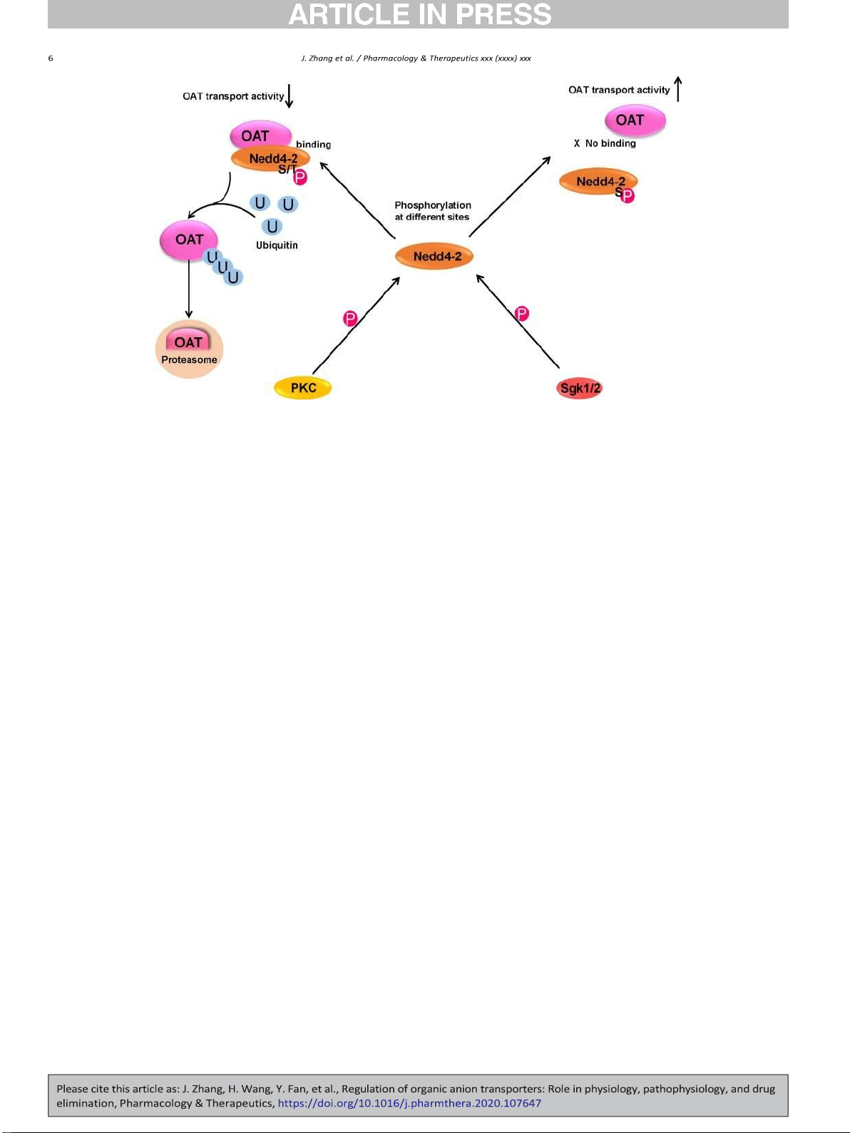
Fig. 2. Phosphorylation of Ubiquitin ligase Nedd4-2 mediates the regulation of OAT transport activity by various kinases. U: ubiquitin, P: Phosphoryl group, S: Serine, T: Threonine, PKC:
Protein kinase C, Sgk1/2: Serum- and glucocorticoid-inducible kinase 1/2, Nedd4-2: Neural precursor cell expressed developmentally down-regulated 4-2.
induces the change in protein conformation, protein activity, cellular
inhibiting OAT activity (H. Wang, Liu, & You, 2018; H. Wang, Xu, Toh,
localization of protein, protein stability, or protein-protein interaction.
Pao, & You, 2016; H. Wang & You, 2017; H. Wang, Zhang, & You,
Many membrane proteins are the substrates of protein kinase-
2019b; J. Zhang, Liu, & You, 2018).
induced direct phosphorylation (Aromolaran, Chahine, & Boutjdir,
Our published and unpublished results indicated PKC activation de-
2018; Mayati et al., 2017). And many protein kinases have been re-
creased OAT expression and transport activity through Nedd4-2 phos-
ported to phosphorylate various transporters. (Cetinkaya et al., 2003;
phorylation instead of directly phosphorylating OAT itself in cultured
Foster & Vaughan, 2017). It is recently demonstrated that Protein Kinase
cells (D. Xu, Wang, & You, 2016a; D. Xu, Wang, Zhang, & You, 2016; D.
A (PKA) activation by Bt2-cAMP induced a significant increase in OAT3
Xu et al., 2017; You et al., 2000; Q. Zhang, Li, Patterson, & You, 2013).
phosphorylation, which was correlated with an enhanced OAT3 trans-
Wolff et al further confirmed such observation by site mutagenesis
port activity in cultured cells. Moreover, Insulin-like growth factor 1
assay: mutagenesis of five canonical PKC phosphorylation sites individ-
(IGF-1), an upstream hormone of PKA signaling, increased OAT3 phos-
ually and in combination resulted in mutants that were insensitive to-
phorylation, and the stimulatory effect was abrogated by H89 (a selec-
ward specific PKC activator dioctanoylglycerol in cultured cells (Wolff
tive PKA inhibitor). IGF-1-stimulated OAT3 phosphorylation was also
et al., 2003). The short-term PKC/Nedd4-2 activation increased OAT
correlated with enhanced OAT3 transport activity and protein expres-
ubiquitination, leading to an accelerated endocytosis of OATs and a re-
sion, and the up-regulation effect was abrogated by PKA inhibitor H89.
duction of its cell surface expression and transport activity in cultured
Therefore, PKA activation by Bt2-cAMP and IGF-1 up-regulated OAT3
cells. In addition, the prolonged PKC/Nedd4-2 activation resulted in
expression and transport activity possibly by directly phosphorylating
the endocytosed OAT to target to proteolytic system for degradation
OAT3 in COS-7 cells (J. Zhang, Yu, & You, 2020).
(Fig. 2). (D. Xu, Wang, & You, 2016a; D. Xu, Wang, Zhang, & You,
Dephosphorylation, countering phosphorylation, refers to a process
2016; D. Xu et al., 2017). Angiotensin II, an endogenous hormone,
which removes phosphoryl group(s) from the target proteins, catalyzed
inhibited OAT1 and OAT3 transport activity through the activation of
by phosphatases. Phosphorylation and dephosphorylation form an op-
PKC/Nedd4-2 pathway (P. Duan, Li, & You, 2010; S. Li, Duan, & You,
posing regulatory network, thus affecting a variety of cellular processes
2009). Nedd4-2 phosphorylation happens not only on serine/threonine
in health and disease (Ardito, Giuliani, Perrone, Troiano, & Lo Muzio,
residues, but also on tyrosine residues. AG490, a specific inhibitor of the
2017; Vitrac, Mallampalli, & Dowhan, 2019). Phosphatase inhibitor
Janus tyrosine kinase 2 (JAK2), reduced OAT3 cell surface expression
okadaic acid inhibited Oat1-mediated transport of para-aminohippurate
and transport activity in cultured cells. The reduced transport activity
(PAH) in cultured cells, which was correlated with an increased phos-
resulted from an enhanced OAT3 ubiquitination, following a reduced
phorylation of Oat1 (You, Kuze, Kohanski, Amsler, & Henderson, 2000).
Nedd4-2 tyrosine phosphorylation and an enhanced interaction be-
tween OAT3 and Nedd4-2. The inhibition effect of AG490 on OAT3
7.2. Regulation of OATs by indirect phosphorylation
was abrogated by knocking down the endogenous Nedd4-2 using
Nedd4-2-specific siRNA (J. Zhang et al., 2018).
Other than directly phosphorylating OATs, protein kinases could also
Some modulators of OATs reduce OAT transport activity through
regulate OATs through phosphorylating OAT-interacting proteins. For
Nedd4-2 phosphorylation, whereas others enhance OAT function
example, Nedd4-2, a ubiquitin ligase, is an OAT-interacting partner.
through phosphorylating Nedd4-2 at different sites. Overexpression of
Ubiquitination of OATs, catalyzed by Nedd4-2, led to the internalization
serum and glucocorticoid-regulated kinase 1 (Sgk1) stimulated OAT3
of OATs from cell surface to intracellular endosomes and subsequent
transport activity in cultured cells. It was shown that Sgk1 phosphory-
degradation. Several protein kinases, hormones, and chemicals excreted
lated Nedd4-2 on Ser327, which weakened the interaction between
their regulation on OATs through phosphorylating Nedd4-2 at different
OAT3 and Nedd4-2, and therefore decreased OAT3 ubiquitination
sites, which either weakened or strengthened the protein-protein inter-
(Fig. 2) (H. Wang & You, 2017). Furthermore, Dexamethasone, an up-
action between OATs and Nedd4-2, and led to either stimulating or
stream hormone of Sgk1, stimulated OAT3 expression and transport
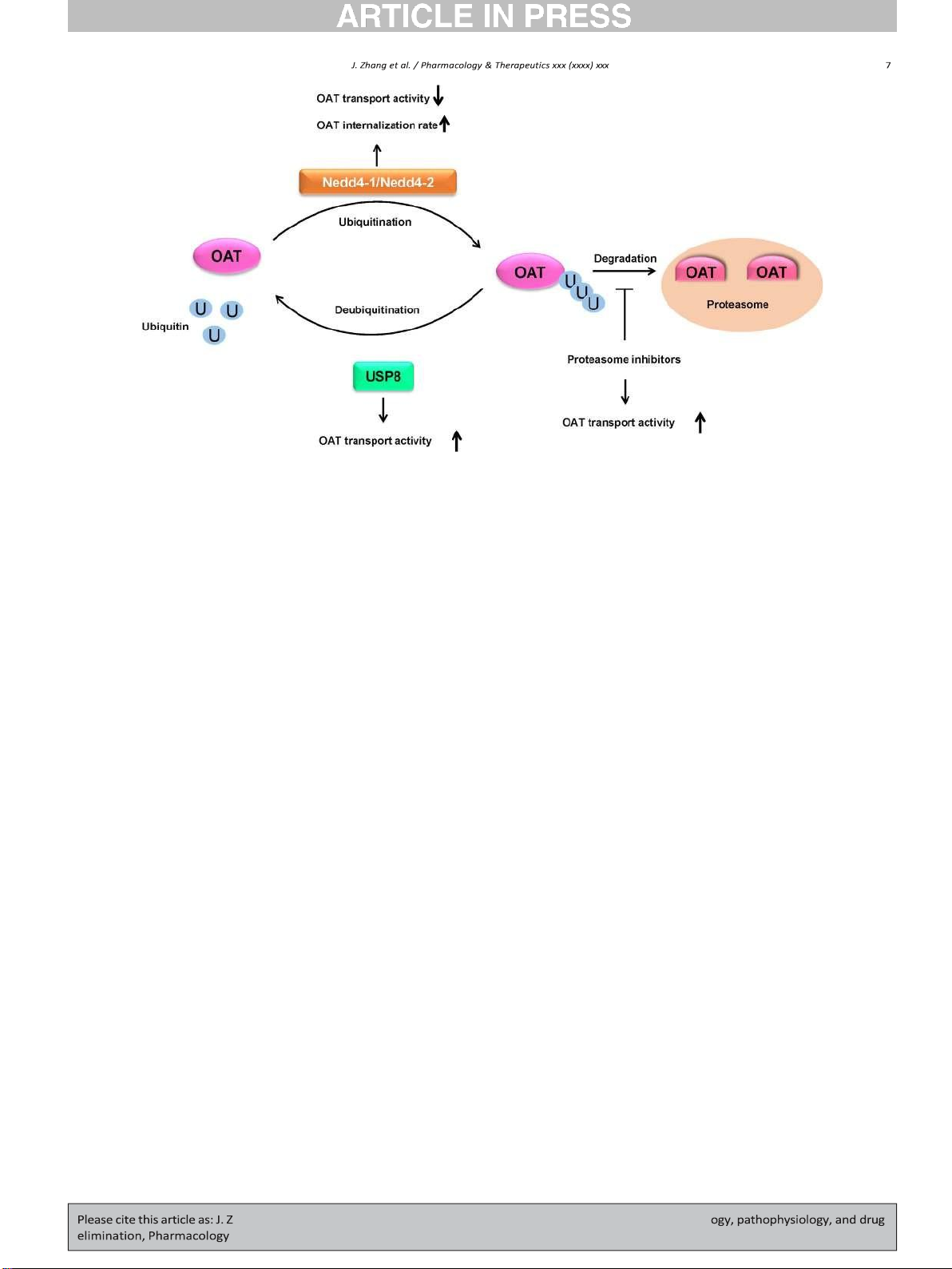
Fig. 3. Regulation of OATs by Ubiquitination, deubiquiting enzyme USP8, and proteasome inhibitors. U: ubiquitin, USP8: ubiquitin-specific proteases 8, Nedd4-1/Nedd4-2: Neural precursor
cell expressed developmentally down-regulated 4-1/4-2.
activity through Nedd4-2 phosphorylation in cultured cells (H. Wang
activity, stability, cellular location, and their interactions with other
et al., 2018). Overexpression of Sgk2, an isoform of Sgk1, enhanced the proteins.
surface expression, total protein expression, and transporter activity
The ubiquitin-proteasome system (UPS) is a major protein degrada-
of OAT1 and OAT4 through impairing the binding between OATs and
tion system. Ubiquitination occurs in a sequence of three enzymatic
Nedd4-2 and decreasing OAT ubiquitination in cultured cells (Fig. 2).
steps and ubiquitinated proteins are targeted to the 26S proteasome
overexpression of Nedd4-2/C821A, a ligase-dead mutant of Nedd4-2,
for degradation (Bence, Sampat, & Kopito, 2001; Gong, Radulovic,
or Nedd4-2 knockdown by Nedd4-2-specific siRNA abrogated the
Figueiredo-Pereira, & Cardozo, 2016; Schwartz & Ciechanover, 2009).
stimulatory effect of Sgk2 on OATs, indicating Sgk2 regulated OATs
The UPS has been reported to be involved in modulating OATs via alter-
possibly through Nedd4-2 phosphorylation (H. Wang et al., 2016; D.
ing cellular location and protein stability (Fig. 3).
Xu, Huang, Toh, & You, 2016). Insulin, an endogenous hormone, in-
creased OAT4 transport activity resulting from an increased OAT4
7.3.1. Regulation of OATs by ubiquitination and deubiquitination
cell surface. Furthermore, insulin up-regulated OAT4 through phos-
Ubiquitination is a PTM that conjugates ubiquitin molecules to target
phorylating Nedd4-2 on Ser327, leading to the impaired association
proteins, catalyzed by ubiquitination enzymes, including ubiquitin-
between OAT4 and Nedd4-2. The up-regulation effect was abrogated
activating enzyme (E1), ubiquitin-conjugating enzyme (E2), and ubiq-
by knocking down the endogenous Nedd4-2 by Nedd4-2-specific
uitin ligase (E3). Nedd4-1 and Nedd4-2, the two E3 ubiquitin ligases,
siRNA (H. Wang et al., 2019b). In summary, the dynamic phosphoryla-
have been identified as important regulators for OATs (Fig. 3). Phorbol
tion of Nedd4-2, a central switch, at different sites exerts opposite reg-
12-Myristate 13-Acetate (PMA), a PKC activator, inhibited OAT trans-
ulations of OATs through different modulators, followed by distinct
port activity and expression in cultured cells. Such inhibition resulted
conformational changes of Nedd4-2 and distinct associations between
from an increased OAT ubiquitination, following PKC-promoted interac-
Nedd4-2 and OATs, which leads to the change in OAT transport activ-
tion between OATs and Nedd4-2, which led to an accelerated internali- ity and expression.
zation of OATs from cell surface to early endosomes and subsequent
degradation (S. Li, Zhang, & You, 2013; D. Xu, Wang, & You, 2016a; Q.
7.3. Regulation of OATs by ubiquitin-proteasome system
Zhang, Suh, Pan, & You, 2012). Overexpression of Nedd4-1 or Nedd4-2
decreased the OAT1 protein expression and transport activity following
Ubiquitin, an 8.6 kDa protein, consists of 76 amino acids. The addi-
the enhanced OAT1 ubiquitination in cultured cells. Knocking down
tion of ubiquitin to the lysine residue(s) of a substrate protein is called
endogenous Nedd4-2 with Nedd4-2-specific siRNA or overexpression
ubiquitination, and ubiquitination could occur in different types of con-
of Nedd4-2/C821A, a ligase-dead mutant of Nedd4-2, abrogated the
jugation including monoubiquitination (conjugation of one single ubiq-
PKC-induced change in OAT1 ubiquitination, expression, and transport
uitin to one single lysine on the substrate), multi-ubiquitination
activity in cultured cells. And PKC-dependent changes in OAT1 ubiquiti-
(conjugation of several monoubiquitin molecules to multiple lysine res-
nation, expression, and transport activity were not affected by knocking
idues on the substrate), and polyubiquitination. Ubiquitin itself has
down endogenous Nedd4-1 or overexpression of Nedd4-1/C867S, a
seven lysine residues and an N-terminal methionine residue including
ligase-dead mutant of Nedd4-1, in cultured cells (D. Xu, Wang, Zhang,
K6, K11, K27, K29, K33, K48, K63, and M1, and a polyubiquitin chain is
& You, 2016). In summary, ubiquitin conjugation to OATs, catalyzed
formed between a glycine residue of one ubiquitin molecule and a ly-
by Nedd4-1 or Nedd4-2, triggers the internalization of OATs from
sine residue or N-terminus of another ubiquitin molecule. The addition
plasma membrane to intracellular endosomes and subsequent degrada-
of a polyubiquitin chain to a single lysine residue of the target protein is
tion in proteolytic systems. As a result, the amount of OATs at the cell
called polyubiquitination (Komander & Rape, 2012; Pickart & Eddins,
surface is reduced, and OAT transport activity is subsequently
2004). Ubiquitination regulates the target proteins by affecting their
decreased. Nedd4-1 is mainly involved in the constitutive OAT
ubiquitination, whereas Nedd4-2 is largely involved in PKC-regulated
to a decrease of OAT1 degradation rate (Fig. 3). Therefore, proteasome OAT ubiquitination.
inhibitors can provide a novel tool to reverse the ubiquitination-
Countering the ubiquitination is a process called deubiquitination
induced downregulation of OATs expression and transport activity, in-
that removes ubiquitin molecules from the target proteins by
dicating their potential influence on the renal OATs-mediated drug dis-
deubiquitinating enzymes (DUBs) (Amerik & Hochstrasser, 2004;
position and drug-drug interactions. Besides, other proteasome
Komander, Clague, & Urbe, 2009). Ubiquitination and deubiquitination
inhibitors are currently in clinical trials, and their influences on the kid-
form an opposing network and are related to a variety of physiological
ney OATs could be attentioned.
and pathological processes (Cai, Culley, Zhao, & Zhao, 2018; Lee et al.,
2006; Y. Wu et al., 2018; Zheng et al., 2016).
7.4. Regulation of OATs by SUMOylation and deSUMOylation
To date, approximately 100 human DUBs has been found, and DUBs
can be classified into six families including the ubiquitin-specific prote-
SUMOylation and deSUMOylation, a pair of opposing and dynamic
ases (USPs), the ubiquitin C-terminal hydrolases (UCHs), the Josephin
PTMs, refer to the process, which adds SUMO to or removes SUMO
family, the ovarian tumor proteases (OTUs), Zn-dependent JAB1/MPN/
from lysine residue of the target protein, catalyzed by specific enzymes.
MOV34 metalloprotease DUBs (JAMMs), and the motif interacting
SUMOylation and deSUMOylation create an on and off switch which is
with ubiquitin (MIU)-containing novel DUB family (MINDYs). (Abdul
essential for biological regulations and are involved in various cellular
Rehman et al., 2016; Clague et al., 2013). A variety of membrane pro-
processes in health and disease (Flotho & Melchior, 2013; C. Guo &
teins, such as channels, receptors, and transporters, are regulated Henley, 2014; Zhao, 2007).
through deubiquitination process by DUBs (Butterworth et al., 2007;
Mines, Goodwin, Limbird, Cui, & Fan, 2009; L. Zhang et al., 2012; R.
7.4.1. Regulation of OATs by SUMOylation Zhou et al., 2013).
SUMOylation is another type of post-translational modification
The investigations on the regulation of OATs by DUBs demonstrated
known as a crucial regulatory mechanism of protein function on both
that overexpression of USP8 decreased OAT1 ubiquitination, leading to
nuclear proteins and cellular membrane proteins (Gareau & Lima,
an increased OAT1 expression at the cell surface and an increased
2010; Gill, 2004; Kang, Saunier, Akhurst, & Derynck, 2008; Plant et al.,
OAT1 transporter activity in cultured cells (Fig. 3). No significant differ-
2010; Rajan et al., 2015; Rajan, Plant, Rabin, Butler, & Goldstein, 2005;
ence in OAT1 expression and transport activity was observed in cells
Ulrich, 2005, 2008). Till now three functional isoforms (SUMO1-3)
transfected with an inactive mutant of USP8 as compared to those in
have been identified in mammals, and all three isoforms are expressed
control cells. Furthermore, knocking down the endogenous USP8 in
in a wide range of tissues, such as brain, lung, liver, Pancreas, and kid-
COS-7 cells by USP8-specific siRNA led to an increase in OAT1
ney. SUMO2 and SUMO3 are usually written as SUMO2/3 since they
ubiquitination which correlated with a reduced OAT1 transport activity
share 97% identity in their amino acid sequences, while SUMO2/3 only
(J. Zhang, Liu, & You, 2017).
shares 50% homology with SUMO1. Although SUMO proteins are con-
sidered as the member of the ubiquitin-like protein family, they only
7.3.2. Regulation of OAT transport activity by proteasome inhibitors
share approximately 18% identity with ubiquitin, and all three SUMO
The ubiquitin-proteasome system is the major proteolytic machin-
proteins are polypeptides of ~12 kDa. Like ubiquitination, the conjuga-
ery that degrades the majority of ubiquitinated intracellular proteins
tion of SUMO to target proteins also involves a series of enzymatic
and certain ubiquitinated membrane proteins in eukaryotic cells
steps. The inactive precursors of SUMO proteins are initially processed
(Alam, Farasyn, Crowe, Ding, & Yue, 2017; Jandial et al., 2009; Ogura
by members of the SUMO1/sentrin specific peptidase (SENP) family to
et al., 2011). Cancer cells have enhanced nuclear factor kappa B
truncate a ten amino acid long fragment from the C terminus, therefore
(NF-kB) activity and are dependent on this signaling pathway for cell
exposing a C-terminal diglycine motif to mature the SUMO proteins.
survival and proliferation. Proteasome inhibition can down-regulate
Then the SUMO-activating enzyme (E1) catalyzes the ATP-dependent
NF-kB-dependent gene expression and lead to an arrest of tumor
formation of a thioester bond between the C terminus of matured
growth. Besides, proteasome inhibition can stabilize tumor suppressor
SUMO and the active cysteine residue of a SUMO-activating enzyme
proteins and repress cell cycle progression. As many cancer cells are
(E1). The activated SUMO is later transferred to a SUMO-conjugating
highly sensitive compared with normal cells for proteasome inhibition,
enzyme (E2). Eventually, SUMO is attached to the specific lysine residue
therefore proteasome has become an important drug target for cancer
on the target protein with the facilitation of SUMO protein ligase (E3). A
therapy (Lenos & Vermeulen, 2016; Thibaudeau & Smith, 2019). The
SUMO substrate can be modified by various types of SUMO conjugation:
26S proteasome is comprised of a proteolytic 20S core particle (20S pro-
monoSUMOylation (conjugation of one single SUMO to one single ly-
teasome) and one or two capped 19S regulatory particles for recogniz-
sine on the target protein), multiSUMOylation (conjugation of several
ing the ubiquitinated proteins (Voges, Zwickl, & Baumeister, 1999).
monoSUMO molecules to multiple lysine residues on the target pro-
Bortezomib, carfilzomib, and ixazomib, approved by FDA for the treat-
tein), and polySUMOylation (extended polySUMO chain). Majority of
ment of patients with multiple myeloma, are reversible or irreversible
SUMO substrates contain the consensus motif, Ψ-K-x-D/E (where ψ is
20S proteasome inhibitors with suppression of chymotrypsin-like activ-
a large hydrophobic residue, K is the lysine conjugated to SUMO, x is
ity. The pharmacodynamic study showed carfilzomib significantly
any amino acid, E is a glutamic acid, and D is an aspartic acid). SUMO2
inhibited the 20S proteasome activity in the kidney of Sprague-
and SUMO3 contain internal SUMO consensus motifs, and therefore
Dawley rats (FDA, 2012). As ubiquitination is one essential post-
are capable of forming polySUMO chains, whereas SUMO1 does not
translational modification which mediates the regulation of OATs, the share such property.
alteration of proteasome activity induced by proteasome inhibitors
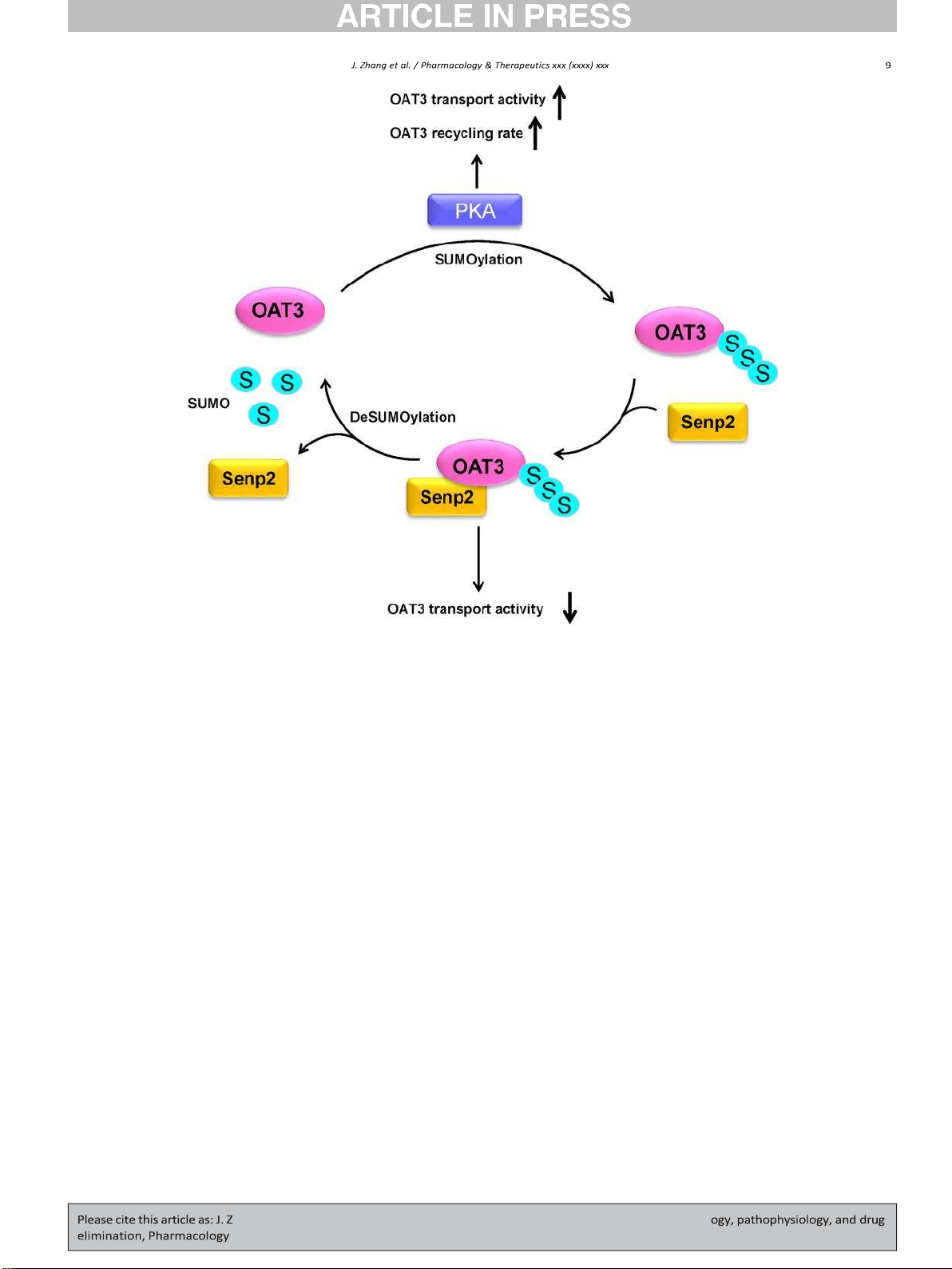
Recently published work showed that in COS-7 cells, OAT3 transport
can also potentially affect the transporter function.
activity and expression at the plasma membrane were increased by
At clinically therapeutic concentrations, incubation of OAT1-
short term PKA activation. Such increase resulted from the enhanced
expressing HEK293 with bortezomib or carfilzomib led to a significant
rate of OAT3 recycling with no change in the rate of OAT3 internaliza-
accumulation of ubiquitinated OAT1, suggesting that ubiquitinated
tion. In addition, OAT3 was identified as a SUMO substrate, and the con-
OAT1 degraded through proteasomes (Fan, Wang, & You, 2018; Fan &
jugation of SUMO2/3 to OAT3 was also PKA-dependent. PKA activation
You, 2020). Bortezomib and carfilzomib significantly stimulated trans-
enhanced OAT3 SUMOylation, and such enhancement can be abrogated
port activity, and there was a positive correlation between the degree
by the presence of PKA-specific inhibitor H-89 (Fig. 4) (H. Wang, Zhang,
of proteasomal inhibition by bortezomib and augmentation of OAT1
& You, 2019a). In Oat3 knockout mice, bile acids such as cholic acid and
transporter activity. Bortezomib- and carfilzomib-induced increase in
taurocholic acid accumulated. Both substances are endogenous sub-
OAT1 surface expression and transport activity were mainly attributed
strates of Oat3 and can activate G protein-coupled receptors (GPCRs)
Fig. 4. Regulation of OATs by SUMOylation and deSUMOylation enzyme Senp2. S: SUMO, Senp2: SUMO1/sentrin specific peptidase 2, PKA: Protein kinase A.
(Deutschmann et al., 2018; Duboc, Tache, & Hofmann, 2014). The acti-
SUMOylation, which paralleled well with an increased OAT3 expression
vation of GPCRs elevated the cAMP level leading to PKA signaling path-
and transport activity. Coimmunoprecipitation experiments revealed
way activation. PKA enhanced OAT3 SUMOylation, recycling rate, and
that Senp2 directly interacted with OAT3/Oat3 both in COS-7 cells and
transport activity. Thus, the accumulation of bile acids contributed by
in rat kidneys (Fig. 4) (Wang & You, 2019).
OAT3 reduction could potentially result in the upregulation of OAT3 ex-
Senps have been reported as key regulators in upholding a balance
pression and function to form a negative feedback loop. This connection
between SUMOylated and unSUMOylated proteins that are crucial for
between OAT endogenous substrates and PTMs of OATs is an interesting
physiological homeostasis. Many investigations indicated the alter-
area to explore. We now know OAT3 is the substrate of SUMOylation.
ations in the amount of Senps under pathophysiological conditions,
However, which lysine residues on OAT3 are responsible for SUMO2/3
and Senps were associated with the progress of a number of diseases,
conjugation is still unknown. Further investigations of mapping the
especially cancer. For example, the level of Senp2 was decreased in
SUMO conjugation sites on OAT3 is needed.
bladder cancer and hepatocellular carcinoma (HCC) tissues, and the
hyperexpression of Senp2 resulted in the suppressions on both bladder
cancer metastasis and HCC development (Shen, Zhu, Yang, & Ji, 2012;
7.4.2. Regulation of OATs by deSUMOylation
Tan et al., 2017). Other than cancers, the overexpression of Senp2
SUMOylation is a dynamic and reversible event, and SUMO is re-
played an important role in the development of congenital heart defects
moved from target protein by SUMO-specific proteases including mem-
and cardiac dysfunction by enhancing deSUMOylation (Kim et al.,
bers of ubiquitin-like specific protease (Ulp, in yeast) and SENP family
2012). Thus, Senps have received increasing recognition as interesting
(in mammals) (Han, Feng, Gu, Li, & Chen, 2018; Hannoun,
targets for drug discovery. 1,2,5-Oxadiazoles were developed as a new
Greenhough, Jaffray, Hay, & Hay, 2010; Miura & Hasegawa, 2010;
class of Senp2 inhibitors, which could have the therapeutic potential
Ulrich, 2005). So far, six human Senp proteins have been isolated and
for many diseases (Kumar, Ito, Takemoto, Yoshida, & Zhang, 2014). Fur-
shown to have the ability to de-conjugate SUMO. Among them (Yeh,
ther studies exploring the effects of Senp2 inhibitors on OAT transport
2009), Senp2 is identified to travel between the nucleus and the cyto-
activity, surface expression, and SUMOylation would be very exciting.
plasm, modulating the activities of some plasma membrane proteins in-
cluding receptors, channels, and transporters (Benson et al., 2007;
Itahana, Yeh, & Zhang, 2006; Qi et al., 2014; Tan et al., 2017).
7.5. Regulation of OATs by Glycosylation
It was recently revealed that in COS-7 cells, overexpression of Senp2,
a member of the SENP family, resulted in a decreased OAT3
Glycosylation, a common and complex PTM of proteins, is the cova-
SUMOylation, which paralleled well with a reduced OAT3 expression
lent attachment of carbohydrates to specific residues of a target protein,
and transport activity. Furthermore, knocking down the endogenous
which expands the proteasome complexity and modulates the target
Senp2 with Senp2-specific siRNA led to an enhanced OAT3
protein via changes in cellular location, protein stability, protein
structure, and protein activity (Eichler, 2019). Glycosylation are classi-
Kolch, & Kholodenko, 2013; Rajan et al., 2015; Yang, Jaffray,
fied into several different protein-sugar linkages, such as N-
Senthinathan, Hay, & Sharrocks, 2003). It would be interesting to ex-
glycosylation, O-glycosylation, C-glycosylation, S-glycosylation, and P-
plore new potential crosstalk among various PTMs of OATs.
glycosylation. N-glycosylation and O-glycosylation are predominantly
found in eukaryotes, and N-glycosylation accounts for more than half
7.7. Remote Sensing and Signaling Hypothesis of OATs
of the protein glycosylation in eukaryotes. N-glycosylation refers to
the attachment of a glycan to the asparagine residue of the target pro-
In the recent years, systems biology studies together with the in-
tein within a consensus peptide sequence (Asn-X-Ser/Thr, X can be
vestigations of OAT knockout mice have indicated that the roles of
any amino acid except proline). For membrane proteins, endoplasmic-
OATs in different organs may form a network, and this network
Golgi pathway, glycosidases, and glycosyltransferases are involved in
allows the intercellular and inter-organ communication. Such com-
the glycosylation process (Christiansen et al., 2014). Many membrane
munication between cells, as well as between organs, regulates the
proteins have been identified as the substrates of glycosylation, and gly-
local and whole-body homeostasis. This hypothesis is called remote
cosylation plays a critical role in regulating the function and activity of
sensing and signaling (Ahn & Nigam, 2009; S. K. Nigam, 2018; S. K.
those membrane proteins (L. B. Li et al., 2004). Nigam et al., 2015).
OAT1 has been reported as the substrate of glycosylation. Asp-39 on
The overlapping of substrate specificities among different OAT iso-
OAT1/Oat1 is crucial for substrate recognition of glycosylation, and gly-
forms, the wide tissue distributions of different OAT isoforms
cosylation is essential for the targeting of OAT1/Oat1 onto the plasma
(e.g., kidney, liver, brain, placenta, retina, olfactory mucosa, etc.), and
membrane (Tanaka, Xu, Zhou, & You, 2004). Furthermore, mutagenesis
the various regulations of OAT expression and function mediated by
of glycosylation sites on OAT4 and treatment of tunicamycin, a glycosyl-
signaling molecules secreted from remote tissues into the body fluid
ation inhibitor, resulted in a non-glycosylated OAT4 and the failure of
contribute to the complicated communication network of OATs
targeting OAT4 onto the plasma membrane in cultured cells. In addition,
(Burckhardt, 2012; Roth, Obaidat, & Hagenbuch, 2012; You, 2002). For
OAT4 expressed in CHO-Lec1 cells, carrying oligosaccharides bearing
example, it was reported that indoxyl sulfate, a gut microbiome-
mannose-rich intermediates, had reduced binding affinity towards the
derived metabolite and endogenous OAT substrate, up-regulated OAT1
substrates compared with OAT4 in CHO wild-type cells, and it was con-
via AhR and EGFR signaling under the control of miR-223 in cultured
cluded that processing of added oligosaccharides from mannose-rich
cells, and the up-regulation on OAT1/Oat1 was to react to the elevated
type to complex type was important for modulating OAT4 substrate
indoxyl sulfate level and to maintain homeostasis through inducing
binding affinity (F. Zhou et al., 2005). Congenital disorders of glycosyla-
renal secretion. This phenomenon was observed in cultured cells, rat
tion (CDG), an ever-expanding disease, are a group of inherited meta-
kidneys, and human kidneys (Jansen et al., 2019). Oat3 was involved
bolic disorders, which affecting glycosylation process. As many steps
in the regulation of blood pressure through the remote sensing and sig-
and enzymes are involved in the glycosylation process, CDG patients
naling. The blood pressure of Oat3 knockout mice was 15% lower than
commonly are deficient of one or more enzymes for one or more glyco-
that of control mice. Metabolomic analysis indicated that plasma con-
sylation steps and show variable clinical symptoms including multi-
centrations of Oat3 substrates were increased in Oat3 knockout mice,
organ dysfunction (Bryant et al., 2020; Ferreira et al., 2018). Kidney dis-
and some endogenous Oat3 substrates could serve as vasodilators, such
function, nephropathy, and lesion of proximal tubule, where OATs are
as thymidine, cAMP, and cGMP, to reduce the blood pressure (Vallon
mainly expressed, are symptoms of CDG, and glycosylation is critical
et al., 2008; Vallon, Eraly, et al., 2008). In addition, the renal excretion
for modulating transport activity of OATs. Thus, OAT function and ex-
of vasodilators, the substrates of Oat3, was reduced responding to the el-
pression in CDG patients would be interesting and exciting to
evated blood pressure, caused by internally and externally environmen- investigate.
tal changes. And then the accumulated vasodilators subsequently
decreased the blood pressure thereby maintaining homeostasis (Ahn &
7.6. Crosstalk between various PTMs Nigam, 2009; Eraly, 2008).
Hormones and growth factors produced and released from the orig-
During the past decade, evidence for comprehensive crosstalk be-
inal organ under the internal and external stimuli arrive at the target
tween different PTM types has stacked up, especially the interplay
organ and regulate the OATs in the target organ through binding to
between the PTMs occurring on the same type of amino acid residue
the receptors and activating the downstream signaling pathways. For
(s). One of the examples is the crosstalk between ubiquitination and
example, Oat3 expression and transport activity were impaired in the
SUMOylation, in which both ubiquitin and SUMO covalently conjugate
streptozotocin-induced type 1 diabetic rats compared with those in
to the lysine residue (s) of a substrate protein. One scenario is that ubiq-
wild-type rats, and insulin treatment abolished the effects.
uitin and SUMO modify the same lysine residue(s) in a target protein
Streptozotocin was used to damage insulin-producing beta cells in the
through competitive manner. On the other hand, SUMO and ubiquitin
pancreas to induce diabetes in animal models. Protein kinase C alpha
may modify different lysine residues in a substrate protein. In such
(PKCα) and phospho-PKCα expression were increased in diabetic rats,
case conjugation of SUMO may potentially mask a nearby ubiquitin con-
and insulin treatment reversed the effects (Phatchawan et al., 2014).
jugation site. Under both circumstances, SUMOylation may preclude the
In addition, insulin stimulated OAT4 expression and transport activity
ubiquitin-mediated degradation of target protein (Hunter & Sun, 2008).
through impairing the interaction between Nedd4-2 and OAT4, and
It was demonstrated that the enhancement of OAT3 SUMOylation by
Nedd4-2 knockdown abolished the stimulation effect of insulin on
PKA activation paralleled with a decrease in OAT3 ubiquitination in cul-
OAT4 in cultured cells (H. Wang et al., 2019b). IGF-1, produced in the
tured cells. Therefore, SUMOylation and ubiquitination may coordi-
liver under stimuli, up-regulated renal OAT3 function through PKA fol-
nately regulate OAT3 through crosstalk (H. Wang et al., 2019a).
lowing binding to its receptor in cultured cells, which was abrogated
Other PTMs, which also occur on lysine residue(s) of the target pro-
by PKA inhibitor H89 and linsitinib (J. Zhang et al., 2020). Besides, it
tein, could potentially be involved in the crosstalk with both
was demonstrated that Angiotensin II, produced in adrenal gland, re-
ubiquitination and SUMOylation. Besides the direct competitions on
duced OAT1 and OAT3 function through PKC in cultured kidney cells
the same lysine residue(s) among these modifications, underlying
(P. Duan et al., 2010; S. Li et al., 2009). Furthermore, parathyroid Hor-
mechanism can be more complex and may not follow a uniform rule
mone, produced in parathyroid glands in the neck, enhanced OAT4 ac-
(Appikonda et al., 2018; Caron, Boyault, & Khochbin, 2005; Gareau &
tivity through a PKA independent pathway in cultured kidney cells (P.
Lima, 2010). In addition, the interplay could also happen between two
Duan, Li, & You, 2012). In remote sensing and signaling model, OATs
or more PTMs modifying different types of amino acid residues
play an essential role in intercellular and inter-organ communication
(Hietakangas et al., 2003; Muller, Matunis, & Dejean, 1998; Nguyen,
and in maintaining local and whole-body homeostasis. Such complex
and dedicated communication is carried out by hormones, small mole-
Burckhardt, G. (2012). Drug transport by organic anion transporters (OATs).
Pharmacology & Therapeutics 136, 106–130. cules and cell signaling.
Bush, K. T., Wu, W., Lun, C., & Nigam, S. K. (2017). The drug transporter OAT3 (SLC22A8)
and endogenous metabolite communication via the gut-liver-kidney axis. The Journal 8. Conclusion
of Biological Chemistry 292, 15789–15803.
Butterworth, M. B., Edinger, R. S., Ovaa, H., Burg, D., Johnson, J. P., & Frizzell, R. A. (2007).
The deubiquitinating enzyme UCH-L3 regulates the apical membrane recycling of the
Elucidating the mechanisms by which OATs are regulated contrib-
epithelial sodium channel. The Journal of Biological Chemistry 282, 37885–37893.
utes significantly to our knowledge of the processes involved in physiol-
Cai, J., Culley, M. K., Zhao, Y., & Zhao, J. (2018). The role of ubiquitination and
ogy, pathology, and drug disposition. It has become increasingly clear
deubiquitination in the regulation of cell junctions. Protein & Cell 9, 754–769.
Caron, C., Boyault, C., & Khochbin, S. (2005). Regulatory cross-talk between lysine acety-
that OATs are regulated by a variety of extracellular factors originated
lation and ubiquitination: Role in the control of protein stability. Bioessays 27,
from different organs, by multiple intracellular signaling pathways, 408–415.
and by various PTMs. Our overview of several important OAT regulators
Cetinkaya, I., Ciarimboli, G., Yalcinkaya, G., Mehrens, T., Velic, A., Hirsch, J. R., ... Schlatter, E.
provides insights into the complexity of this process. Most studies have
(2003). Regulation of human organic cation transporter hOCT2 by PKA, PI3K, and
calmodulin-dependent kinases. American Journal of Physiology. Renal Physiology 284,
examined these regulatory factors in isolation. How they work in con- F293–F302.
cert to modulate OAT function, to improve OAT-related medical treat-
Cha, S. H., Sekine, T., Kusuhara, H., Yu, E., Kim, J. Y., Kim, D. K., ... Endou, H. (2000). Molec-
ment, and to keep body homeostasis are important questions that
ular cloning and characterization of multispecific organic anion transporter 4
expressed in the placenta. The Journal of Biological Chemistry 275, 4507–4512. continue to be addressed.
Chen, J., Terada, T., Ogasawara, K., Katsura, T., & Inui, K. (2008). Adaptive responses of
renal organic anion transporter 3 (OAT3) during cholestasis. American Journal of Phys-
Declaration of Competing Interest
iology. Renal Physiology 295, F247–F252.
Cheng, Q., Aleksunes, L. M., Manautou, J. E., Cherrington, N. J., Scheffer, G. L., Yamasaki, H.,
& Slitt, A. L. (2008). Drug-metabolizing enzyme and transporter expression in a
The authors declare that there are no conflicts of interest.
mouse model of diabetes and obesity. Molecular Pharmaceutics 5, 77–91.
Chioukh, R., Noel-Hudson, M. S., Ribes, S., Fournier, N., Becquemont, L., & Verstuyft, C. Acknowledgements
(2014). Proton pump inhibitors inhibit methotrexate transport by renal basolateral
organic anion transporter hOAT3. Drug Metabolism and Disposition 42, 2041–2048.
Cho, S. K., Kim, S., Chung, J. Y., & Jee, S. H. (2015). Discovery of URAT1 SNPs and association
This work was supported by grants (to Dr. Guofeng You) from Na-
between serum uric acid levels and URAT1. BMJ Open 5, e009360.
tional Institute of General Medical Sciences (R01-GM079123, R01-
Christiansen, M. N., Chik, J., Lee, L., Anugraham, M., Abrahams, J. L., & Packer, N. H. (2014).
Cell surface protein glycosylation in cancer. Proteomics 14, 525–546. GM097000 and R01-GM127788).
Clague, M. J., Barsukov, I., Coulson, J. M., Liu, H., Rigden, D. J., & Urbe, S. (2013).
Deubiquitylases from genes to organism. Physiological Reviews 93, 1289–1315. Reference
Cutler, M. J., Urquhart, B. L., Velenosi, T. J., Meyer Zu Schwabedissen, H. E., Dresser, G. K.,
Leake, B. F., ... Freeman, D. J. (2012). In vitro and in vivo assessment of renal drug
Abdul Rehman, S. A., Kristariyanto, Y. A., Choi, S. Y., Nkosi, P. J., Weidlich, S., Labib, K., ...
transporters in the disposition of mesna and dimesna. Journal of Clinical
Kulathu, Y. (2016). MINDY-1 Is a member of an evolutionarily conserved and struc-
Pharmacology 52, 530–542.
turally distinct new family of deubiquitinating enzymes. Molecular Cell 63, 146–155.
Czuba, L. C., Hillgren, K. M., & Swaan, P. W. (2018). Post-translational modifications of
Ahn, S. Y., & Nigam, S. K. (2009). Toward a systems level understanding of organic anion
transporters. Pharmacology & Therapeutics 192, 88–99.
and other multispecific drug transporters: A remote sensing and signaling hypothe-
Dantzler, W. H., & Wright, S. H. (2003). The molecular and cellular physiology of
sis. Molecular Pharmacology 76, 481–490.
basolateral organic anion transport in mammalian renal tubules. Biochimica et
Alam, K., Farasyn, T., Crowe, A., Ding, K., & Yue, W. (2017). Treatment with proteasome
Biophysica Acta 1618, 185–193.
inhibitor bortezomib decreases organic anion transporting polypeptide (OATP)
Deutschmann, K., Reich, M., Klindt, C., Droge, C., Spomer, L., Haussinger, D., & Keitel, V.
1B3-mediated transport in a substrate-dependent manner. PLoS One 12, e0186924.
(2018). Bile acid receptors in the biliary tree: TGR5 in physiology and disease.
Amerik, A. Y., & Hochstrasser, M. (2004). Mechanism and function of deubiquitinating en-
Biochimica et Biophysica Acta - Molecular Basis of Disease 1864, 1319–1325.
zymes. Biochimica et Biophysica Acta 1695, 189–207.
Duan, G., & Walther, D. (2015). The roles of post-translational modifications in the con-
Anzai, N., Kanai, Y., & Endou, H. (2006). Organic anion transporter family: current knowl-
text of protein interaction networks. PLoS Computational Biology 11, e1004049.
edge. Journal of Pharmacological Sciences 100, 411–426.
Duan, P., Li, S., & You, G. (2010). Angiotensin II inhibits activity of human organic anion
Appikonda, S., Thakkar, K. N., Shah, P. K., Dent, S. Y. R., Andersen, J. N., & Barton, M. C.
transporter 3 through activation of protein kinase Calpha: accelerating endocytosis
(2018). Cross-talk between chromatin acetylation and SUMOylation of tripartite
of the transporter. European Journal of Pharmacology 627, 49–55.
motif-containing protein 24 (TRIM24) impacts cell adhesion. The Journal of
Duan, P., Li, S., & You, G. (2011). Transmembrane peptide as potent inhibitor of oligomer-
Biological Chemistry 293, 7476–7485.
ization and function of human organic anion transporter 1. Molecular Pharmacology
Ardito, F., Giuliani, M., Perrone, D., Troiano, G., & Lo Muzio, L. (2017). The crucial role of 79, 569–574.
protein phosphorylation in cell signaling and its use as targeted therapy (Review).
Duan, P., Li, S., & You, G. (2012). Regulation of human organic anion transporter 4 by para-
International Journal of Molecular Medicine 40, 271–280.
thyroid hormone-related protein and protein kinase A. International Journal of
Aromolaran, A. S., Chahine, M., & Boutjdir, M. (2018). Regulation of cardiac voltage-gated
Biochemistry and Molecular Biology 3, 322–327.
sodium channel by kinases: Roles of protein kinases A and C. Handbook of
Duan, P., & You, G. (2010). Short-term regulation of organic anion transporters.
Experimental Pharmacology 246, 161–184.
Pharmacology & Therapeutics 125, 55–61.
Baliga, R., Ueda, N., Walker, P. D., & Shah, S. V. (1999). Oxidant mechanisms in toxic acute
Duboc, H., Tache, Y., & Hofmann, A. F. (2014). The bile acid TGR5 membrane receptor:
renal failure. Drug Metabolism Reviews 31, 971–997.
from basic research to clinical application. Digestive and Liver Disease 46, 302–312.
Bence, N. F., Sampat, R. M., & Kopito, R. R. (2001). Impairment of the ubiquitin-
Eichler, J. (2019). Protein glycosylation. Current Biology 29, R229–R231.
proteasome system by protein aggregation. Science 292, 1552–1555.
Emami Riedmaier, A., Nies, A. T., Schaeffeler, E., & Schwab, M. (2012). Organic anion trans-
Benson, M. D., Li, Q. J., Kieckhafer, K., Dudek, D., Whorton, M. R., Sunahara, R. K., ... Martens,
porters and their implications in pharmacotherapy. Pharmacological Reviews 64,
J. R. (2007). SUMO modification regulates inactivation of the voltage-gated potassium 421–449.
channel Kv1.5. Proceedings of the National Academy of Sciences of the United States of
Eraly, S. A. (2008). Organic anion transporter 3 inhibitors as potential novel antihyperten-
America 104, 1805–1810.
sives. Pharmacological Research 58, 257–261.
Bhatnagar, V., Richard, E. L., Wu, W., Nievergelt, C. M., Lipkowitz, M. S., Jeff, J., ... Nigam, S.
Eraly, S. A., Vallon, V., Rieg, T., Gangoiti, J. A., Wikoff, W. R., Siuzdak, G., ... Nigam, S. K.
K. (2016). Analysis of ABCG2 and other urate transporters in uric acid homeostasis in
(2008). Multiple organic anion transporters contribute to net renal excretion of uric
chronic kidney disease: potential role of remote sensing and signaling. Clinical Kidney
acid. Physiological Genomics 33, 180–192. Journal 9, 444–453.
Eraly, S. A., Vallon, V., Vaughn, D. A., Gangoiti, J. A., Richter, K., Nagle, M., ... Nigam, S. K.
Bischoff, A., Bucher, M., Gekle, M., & Sauvant, C. (2014a). Differential effect of COX1 and
(2006). Decreased renal organic anion secretion and plasma accumulation of endog-
COX2 inhibitors on renal outcomes following ischemic acute kidney injury.
enous organic anions in OAT1 knock-out mice. The Journal of Biological Chemistry 281,
American Journal of Nephrology 40, 1–11. 5072–5083.
Bischoff, A., Bucher, M., Gekle, M., & Sauvant, C. (2014b). PAH clearance after renal ische-
Fan, Y., Wang, G., & You, G. (2018). Proteasome inhibitors bortezomib and carfilzomib
mia and reperfusion is a function of impaired expression of basolateral Oat1 and
stimulate the transport activity of human organic anion transporter 3 (abstract).
Oat3. Physiological Reports 2, e00243.
American Association of Pharmaceutical Scientists Annual Meeting, Washington, DC., Ab-
Brandoni, A., Anzai, N., Kanai, Y., Endou, H., & Torres, A. M. (2006). Renal elimination of p-
stract No. W1030-05-038.
aminohippurate (PAH) in response to three days of biliary obstruction in the rat. The
Fan, Y., & You, G. (2020). Proteasome inhibitors bortezomib and carfilzomib stimulate the
role of OAT1 and OAT3. Biochimica et Biophysica Acta 1762, 673–682.
transport activity of human organic anion transporter 1. Molecular Pharmacology 97
Bryant, E. M., Millichap, J. J., Spinelli, E., Calhoun, J. D., Miller, C., Giannelli, J., ... Charrow, J. (6), 384–391.
(2020). Oligosaccharyltransferase complex-congenital disorders of glycosylation: A
FDA (2012). Carfilzomib Pharmacology Review(S). APPLICATION NUMBER: 202714Orig1s
novel congenital disorder of glycosylation. American Journal of Medical Genetics. Part
000. https://www.accessdata.fda.gov/drugsatfda_docs/nda/2012/202714Orig202711 A 182A, 1460–1465. s202000PharmR.pdf.
Feng, Y., Wang, C., Liu, Q., Meng, Q., Huo, X., Liu, Z., ... Liu, K. (2016). Bezafibrate-
Kikuchi, R., Kusuhara, H., Hattori, N., Kim, I., Shiota, K., Gonzalez, F. J., & Sugiyama, Y.
mizoribine interaction: Involvement of organic anion transporters OAT1 and OAT3
(2007). Regulation of tissue-specific expression of the human and mouse urate trans-
in rats. European Journal of Pharmaceutical Sciences 81, 119–128.
porter 1 gene by hepatocyte nuclear factor 1 alpha/beta and DNA methylation.
Ferreira, C. R., Altassan, R., Marques-Da-Silva, D., Francisco, R., Jaeken, J., & Morava, E.
Molecular Pharmacology 72, 1619–1625.
(2018). Recognizable phenotypes in CDG. Journal of Inherited Metabolic Disease 41,
Kikuchi, R., Kusuhara, H., Hattori, N., Shiota, K., Kim, I., Gonzalez, F. J., & Sugiyama, Y. 541–553.
(2006). Regulation of the expression of human organic anion transporter 3 by hepa-
Flotho, A., & Melchior, F. (2013). Sumoylation: a regulatory protein modification in health
tocyte nuclear factor 1alpha/beta and DNA methylation. Molecular Pharmacology 70,
and disease. Annual Review of Biochemistry 82, 357–385. 887–896.
Foster, J. D., & Vaughan, R. A. (2017). Phosphorylation mechanisms in dopamine trans-
Kim, E. Y., Chen, L., Ma, Y., Yu, W., Chang, J., Moskowitz, I. P., & Wang, J. (2012). Enhanced
porter regulation. Journal of Chemical Neuroanatomy 83-84, 10–18.
desumoylation in murine hearts by overexpressed SENP2 leads to congenital heart
Fujita, T., Ishihara, K., Yasuda, S., Nakamura, T., Maeda, M., Kobayashi, M., ... Majima, M.
defects and cardiac dysfunction. Journal of Molecular and Cellular Cardiology 52,
(2012). In vivo kinetics of indoxyl sulfate in humans and its renal interaction with 638–649.
angiotensin-converting enzyme inhibitor quinapril in rats. The Journal of
Koepsell, H. (2013). The SLC22 family with transporters of organic cations, anions and
Pharmacology and Experimental Therapeutics 341, 626–633.
zwitterions. Molecular Aspects of Medicine 34, 413–435.
Gareau, J. R., & Lima, C. D. (2010). The SUMO pathway: emerging mechanisms that shape
Kolz, M., Johnson, T., Sanna, S., Teumer, A., Vitart, V., Perola, M., ... Gieger, C. (2009). Meta-
specificity, conjugation and recognition. Nature Reviews. Molecular Cell Biology 11,
analysis of 28,141 individuals identifies common variants within five new loci that 861–871.
influence uric acid concentrations. PLoS Genetics e1000504, 5.
Giacomini, K. M., Huang, S. M., Tweedie, D. J., Benet, L. Z., Brouwer, K. L., Chu, X., ...
Komander, D., Clague, M. J., & Urbe, S. (2009). Breaking the chains: structure and function
Consortium, I. T. (2010). Membrane transporters in drug development. Nature
of the deubiquitinases. Nature Reviews. Molecular Cell Biology 10, 550–563.
Reviews. Drug Discovery 9, 215–236.
Komander, D., & Rape, M. (2012). The ubiquitin code. Annual Review of Biochemistry 81,
Gibaldi, M., & Schwartz, M. A. (1968). Apparent effect of probenecid on the distribution of 203–229.
penicillins in man. Clinical Pharmacology and Therapeutics 9, 345–349.
Komazawa, H., Yamaguchi, H., Hidaka, K., Ogura, J., Kobayashi, M., & Iseki, K. (2013). Renal
Gill, G. (2004). SUMO and ubiquitin in the nucleus: different functions, similar mecha-
uptake of substrates for organic anion transporters Oat1 and Oat3 and organic cation
nisms? Genes & Development 18, 2046–2059.
transporters Oct1 and Oct2 is altered in rats with adenine-induced chronic renal fail-
Gong, B., Radulovic, M., Figueiredo-Pereira, M. E., & Cardozo, C. (2016). The ubiquitin-
ure. Journal of Pharmaceutical Sciences 102, 1086–1094.
proteasome system: Potential therapeutic targets for Alzheimer’s disease and spinal
Kong, F., Pang, X., Zhong, K., Guo, Z., Li, X., Zhong, D., & Chen, X. (2017). Increased plasma
cord injury. Frontiers in Molecular Neuroscience 9, 4.
exposures of conjugated metabolites of morinidazole in renal failure patients: A crit-
Guo, C., & Henley, J. M. (2014). Wrestling with stress: Roles of protein SUMOylation and
ical role of uremic toxins. Drug Metabolism and Disposition 45, 593–603.
deSUMOylation in cell stress response. IUBMB Life 66, 71–77.
Kozaki, T., Tagashira, M., Yamanishi, K., Ellis, B., Kayanoki, T., Ooishi, R., ... Tsurui, K. (2015).
Guo, X., Meng, Q., Liu, Q., Wang, C., Sun, H., Peng, J., ... Liu, K. (2013). JBP485 improves
Evaluation of drug-drug interaction between the novel cPLA2 inhibitor AK106-
gentamicin-induced acute renal failure by regulating the expression and function of
001616 and methotrexate in rheumatoid arthritis patients. Xenobiotica 45, 615–624.
Oat1 and Oat3 in rats. Toxicology and Applied Pharmacology 271, 285–295.
Kumar, A., Ito, A., Takemoto, M., Yoshida, M., & Zhang, K. Y. (2014). Identification of 1,2,5-
Han, Z. J., Feng, Y. H., Gu, B. H., Li, Y. M., & Chen, H. (2018). The post-translational modifi-
oxadiazoles as a new class of SENP2 inhibitors using structure based virtual screen-
cation, SUMOylation, and cancer (Review). International Journal of Oncology 52,
ing. Journal of Chemical Information and Modeling 54, 870–880. 1081–1094.
Lee, H. J., Kim, M. S., Shin, J. M., Park, T. J., Chung, H. M., & Baek, K. H. (2006). The expres-
Hannoun, Z., Greenhough, S., Jaffray, E., Hay, R. T., & Hay, D. C. (2010). Post-translational
sion patterns of deubiquitinating enzymes, USP22 and Usp22. Gene Expression
modification by SUMO. Toxicology 278, 288–293. Patterns 6, 277–284.
He, J. B., Ren, Y. L., Sun, Q. S., You, G. Y., Zhang, L., Zou, P., ... He, H. W. (2014). Design, syn-
Lenos, K. J., & Vermeulen, L. (2016). Cancer stem cells don’t waste their time cleaning-low
thesis and molecular docking of amide and urea derivatives as Escherichia coli PDHc-
proteasome activity, a marker for cancer stem cell function. Annals of Translational
E1 inhibitors. Bioorganic & Medicinal Chemistry 22, 3180–3186. Medicine 4, 519.
Hietakangas, V., Ahlskog, J. K., Jakobsson, A. M., Hellesuo, M., Sahlberg, N. M., Holmberg, C.
Li, L. B., Chen, N., Ramamoorthy, S., Chi, L., Cui, X. N., Wang, L. C., & Reith, M. E. (2004). The
I., ... Sistonen, L. (2003). Phosphorylation of serine 303 is a prerequisite for the stress-
role of N-glycosylation in function and surface trafficking of the human dopamine
inducible SUMO modification of heat shock factor 1. Molecular and Cellular Biology 23,
transporter. The Journal of Biological Chemistry 279, 21012–21020. 2953–2968.
Li, S., Duan, P., & You, G. (2009). Regulation of human organic anion transporter 1 by ANG
Hillgren, K. M., Keppler, D., Zur, A. A., Giacomini, K. M., Stieger, B., Cass, C. E., ... Consortium,
II: involvement of protein kinase Calpha. American Journal of Physiology. Endocrinol-
I. T. (2013). Emerging transporters of clinical importance: an update from the Inter-
ogy and Metabolism 296, E378–E383.
national Transporter Consortium. Clinical Pharmacology and Therapeutics 94, 52–63.
Li, S., Zhang, Q., & You, G. (2013). Three ubiquitination sites of organic anion transporter-1
Hong, M., Li, S., Zhou, F., Thomas, P. E., & You, G. (2010). Putative transmembrane domain
synergistically mediate protein kinase C-dependent endocytosis of the transporter.
12 of the human organic anion transporter hOAT1 determines transporter stability
Molecular Pharmacology 84, 139–146.
and maturation efficiency. The Journal of Pharmacology and Experimental
Lin, S. P., Yu, C. P., Hou, Y. C., Huang, C. Y., Ho, L. C., & Chan, S. L. (2018). Transporter-
Therapeutics 332, 650–658.
mediated interaction of indican and methotrexate in rats. Journal of Food and Drug
Hong, M., Xu, W., Yoshida, T., Tanaka, K., Wolff, D. J., Zhou, F., ... You, G. (2005). Human or-
Analysis 26, S133–S140.
ganic anion transporter hOAT1 forms homooligomers. The Journal of Biological
Lipkowitz, M. S. (2012). Regulation of uric acid excretion by the kidney. Current
Chemistry 280, 32285–32290.
Rheumatology Reports 14, 179–188.
Hsueh, C. H., Yoshida, K., Zhao, P., Meyer, T. W., Zhang, L., Huang, S. M., & Giacomini, K. M. (2016). Identi
Liu, C., Zhang, J., & You, G. (2019). Interaction of anticancer drugs with human organic
fication and quantitative assessment of uremic solutes as inhibitors of
anion transporter hOAT4. Journal of Oncology 2019, 1951786.
renal organic anion transporters, OAT1 and OAT3. Molecular Pharmaceutics 13, 3130–3140.
Liu, T., Guo, X., Meng, Q., Wang, C., Liu, Q., Sun, H., ... Liu, K. (2012). Effect of JBP485 on ob-
Hunter, T., & Sun, H. (2008). Crosstalk between the SUMO and ubiquitin pathways. Ernst
structive jaundice is related to regulation of renal Oat1, Oat3 and Mrp2 expression in
Schering Foundation Symposium Proceedings, 1–16.
ANIT-treated rats. Peptides 36, 78–85.
Huo, X., & Liu, K. (2018). Renal organic anion transporters in drug-drug interactions and
Lozano, E., Briz, O., Macias, R. I. R., Serrano, M. A., Marin, J. J. G., & Herraez, E. (2018).
diseases. European Journal of Pharmaceutical Sciences 112, 8–19.
Journal of Personalized Medicine, 8.
Ichida, K., Matsuo, H., Takada, T., Nakayama, A., Murakami, K., Shimizu, T., ... Suzuki, H.
Maeda, K., Tian, Y., Fujita, T., Ikeda, Y., Kumagai, Y., Kondo, T., ... Sugiyama, Y. (2014).
(2012). Decreased extra-renal urate excretion is a common cause of hyperuricemia.
Inhibitory effects of p-aminohippurate and probenecid on the renal clearance of
Nature Communications 3, 764.
adefovir and benzylpenicillin as probe drugs for organic anion transporter (OAT) 1
Ikemura, K., Hamada, Y., Kaya, C., Enokiya, T., Muraki, Y., Nakahara, H., ... Okuda, M.
and OAT3 in humans. European Journal of Pharmaceutical Sciences 59, 94–103.
(2016). Lansoprazole exacerbates pemetrexed-mediated hematologic toxicity by
Maher, J. M., Slitt, A. L., Callaghan, T. N., Cheng, X., Cheung, C., Gonzalez, F. J., & Klaassen, C.
competitive inhibition of renal basolateral human organic anion transporter 3. Drug
D. (2006). Alterations in transporter expression in liver, kidney, and duodenum after
Metabolism and Disposition 44, 1543–1549.
targeted disruption of the transcription factor HNF1alpha. Biochemical Pharmacology
Itahana, Y., Yeh, E. T., & Zhang, Y. (2006). Nucleocytoplasmic shuttling modulates activity 72, 512–522.
and ubiquitination-dependent turnover of SUMO-specific protease 2. Molecular and
Martovetsky, G., Tee, J. B., & Nigam, S. K. (2013). Hepatocyte nuclear factors 4alpha and
Cellular Biology 26, 4675–4689.
1alpha regulate kidney developmental expression of drug-metabolizing enzymes
Jandial, D. D., Farshchi-Heydari, S., Larson, C. A., Elliott, G. I., Wrasidlo, W. J., & Howell, S. B.
and drug transporters. Molecular Pharmacology 84, 808–823.
(2009). Enhanced delivery of cisplatin to intraperitoneal ovarian carcinomas medi-
Mathialagan, S., Piotrowski, M. A., Tess, D. A., Feng, B., Litchfield, J., & Varma, M. V. (2017).
ated by the effects of bortezomib on the human copper transporter 1. Clinical
Quantitative prediction of human renal clearance and drug-drug interactions of or-
Cancer Research 15, 553–560.
ganic anion transporter substrates using in vitro transport data: A relative activity
Jansen, J., Jansen, K., Neven, E., Poesen, R., Othman, A., van Mil, A., ... Masereeuw, R. (2019).
factor approach. Drug Metabolism and Disposition 45, 409–417.
Remote sensing and signaling in kidney proximal tubules stimulates gut
Mayati, A., Moreau, A., Le Vee, M., Stieger, B., Denizot, C., Parmentier, Y., & Fardel, O.
microbiome-derived organic anion secretion. Proceedings of the National Academy of
(2017). Protein kinases C-mediated regulations of drug transporter activity, localiza-
Sciences of the United States of America 116, 16105–16110.
tion and expression. International Journal of Molecular Sciences 18.
Jia, Y., Liu, Z., Wang, C., Meng, Q., Huo, X., Liu, Q., ... Liu, K. (2016). P-gp, MRP2 and OAT1/
Mines, M. A., Goodwin, J. S., Limbird, L. E., Cui, F. F., & Fan, G. H. (2009). Deubiquitination of
OAT3 mediate the drug-drug interaction between resveratrol and methotrexate.
CXCR4 by USP14 is critical for both CXCL12-induced CXCR4 degradation and chemo-
Toxicology and Applied Pharmacology 306, 27–35.
taxis but not ERK ativation. The Journal of Biological Chemistry 284, 5742–5752.
Kang, J. S., Saunier, E. F., Akhurst, R. J., & Derynck, R. (2008). The type I TGF-beta receptor is
Miura, K., & Hasegawa, P. M. (2010). Sumoylation and other ubiquitin-like post-
covalently modified and regulated by sumoylation. Nature Cell Biology 10, 654–664.
translational modifications in plants. Trends in Cell Biology 20, 223–232.
Motohashi, H., Nakao, Y., Masuda, S., Katsura, T., Kamba, T., Ogawa, O., & Inui, K. (2013).
nuclear factor 1 alpha/beta. The Journal of Pharmacology and Experimental
Precise comparison of protein localization among OCT, OAT, and MATE in human kid-
Therapeutics 324, 784–790.
ney. Journal of Pharmaceutical Sciences 102, 3302–3308.
Sakiyama, M., Matsuo, H., Shimizu, S., Nakashima, H., Nakayama, A., Chiba, T., ...
Muller, S., Matunis, M. J., & Dejean, A. (1998). Conjugation with the ubiquitin-related
Shinomiya, N. (2014). A common variant of organic anion transporter 4 (OAT4/
modifier SUMO-1 regulates the partitioning of PML within the nucleus. The EMBO
SLC22A11) gene is associated with renal underexcretion type gout. Drug Journal 17, 61–70.
Metabolism and Pharmacokinetics 29, 208–210.
Narumi, K., Sato, Y., Kobayashi, M., Furugen, A., Kasashi, K., Yamada, T., ... Iseki, K. (2017).
Sakurai, Y., Motohashi, H., Ueo, H., Masuda, S., Saito, H., Okuda, M., ... Inui, K. (2004). Ex-
Effects of proton pump inhibitors and famotidine on elimination of plasma metho-
pression levels of renal organic anion transporters (OATs) and their correlation
trexate: Evaluation of drug-drug interactions mediated by organic anion transporter
with anionic drug excretion in patients with renal diseases. Pharmaceutical Research
3. Biopharmaceutics & Drug Disposition 38, 501–508. 21, 61–67.
Naud, J., Michaud, J., Beauchemin, S., Hebert, M. J., Roger, M., Lefrancois, S., ... Pichette, V.
Schneider, R., Meusel, M., Betz, B., Held, C., Möller-Ehrlich, K., Büttner-Herold, M., ...
(2011). Effects of chronic renal failure on kidney drug transporters and cytochrome
Sauvant, C. (2015). Oat1/3 restoration protects against renal damage after ischemic
P450 in rats. Drug Metabolism and Disposition 39, 1363–1369.
AKI. American Journal of Physiology. Renal Physiology 308, F198–F208.
Nguyen, L. K., Kolch, W., & Kholodenko, B. N. (2013). When ubiquitination meets phos-
Schwartz, A. L., & Ciechanover, A. (2009). Targeting proteins for destruction by the ubiq-
phorylation: a systems biology perspective of EGFR/MAPK signalling. Cell
uitin system: implications for human pathobiology. Annual Review of Pharmacology
Communication and Signaling: CCS 11, 52.
and Toxicology 49, 73–96.
Nigam, A. K., Li, J. G., Lall, K., Shi, D., Bush, K. T., Bhatnagar, V., ... Nigam, S. K. (2020). Unique
Schwenk, M. H., & Pai, A. B. (2016). Drug transporter function–implications in CKD.
metabolite preferences of the drug transporters OAT1 and OAT3 analyzed by ma -
Advances in Chronic Kidney Disease 23, 76–81.
chine learning. The Journal of Biological Chemistry 295, 1829–1842.
Shen, H. J., Zhu, H. Y., Yang, C., & Ji, F. (2012). SENP2 regulates hepatocellular carcinoma
Nigam, S. K. (2018). The SLC22 transporter family: A paradigm for the impact of drug
cell growth by modulating the stability of beta-catenin. Asian Pacific Journal of Cancer
transporters on metabolic pathways, signaling, and disease. Annual Review of
Prevention 13, 3583–3587.
Pharmacology and Toxicology 58, 663–687.
Spoel, S. H. (2018). Orchestrating the proteome with post-translational modifications.
Nigam, S. K., Bush, K. T., Martovetsky, G., Ahn, S. Y., Liu, H. C., Richard, E., ... Wu, W. (2015).
Journal of Experimental Botany 69, 4499–4503.
The organic anion transporter (OAT) family: a systems biology perspective.
Srimaroeng, C., Perry, J. L., & Pritchard, J. B. (2008). Physiology, structure, and regulation of
Physiological Reviews 95, 83–123.
the cloned organic anion transporters. Xenobiotica 38, 889–935.
Ogasawara, K., Terada, T., Asaka, J., Katsura, T., & Inui, K. (2006). Human organic anion
transporter 3 gene is regulated constitutively and inducibly via a cAMP-response el-
Sun, C. Y., Wu, M. S., Lee, C. C., Chen, S. H., Lo, K. C., & Chen, Y. H. (2018). A novel SNP in the
ement. The Journal of Pharmacology and Experimental Therapeutics 319, 317–322.
5’ regulatory region of organic anion transporter 1 is associated with chronic kidney
Ogasawara, K., Terada, T., Asaka, J., Katsura, T., & Inui, K. (2007). Hepatocyte nuclear
disease. Scientific Reports 8, 8085.
factor-4{alpha} regulates the human organic anion transporter 1 gene in the kidney.
Takahara, N., Saga, T., Inubushi, M., Kusuhara, H., Seki, C., Ito, S., ... Fujibayashi, Y. (2013).
American Journal of Physiology. Renal Physiology 292, F1819–F1826.
Drugs interacting with organic anion transporter-1 affect uptake of Tc-99m-
Ogura, M., Ayaori, M., Terao, Y., Hisada, T., Iizuka, M., Takiguchi, S., ... Ikewaki, K. (2011).
mercaptoacetyl-triglycine (MAG3) in the human kidney: therapeutic drug interac-
Proteasomal inhibition promotes ATP-binding cassette transporter A1 (ABCA1) and
tion in Tc-99m-MAG3 diagnosis of renal function and possible application of Tc-
ABCG1 expression and cholesterol efflux from macrophages in vitro and in vivo.
99m-MAG3 for drug development. Nuclear Medicine and Biology 40, 643–650.
Arteriosclerosis, Thrombosis, and Vascular Biology 31, 1980–1987.
Taki, K., Nakamura, S., Miglinas, M., Enomoto, A., & Niwa, T. (2006). Accumulation of in-
Pannu, N., & Nadim, M. K. (2008). An overview of drug-induced acute kidney injury.
doxyl sulfate in OAT1/3-positive tubular cells in kidneys of patients with chronic
Critical Care Medicine 36, S216–S223.
renal failure. Journal of Renal Nutrition 16, 199–203.
Parvez, M. M., Kaisar, N., Shin, H. J., Jung, J. A., & Shin, J. G. (2016). Inhibitory interaction
Tan, M., Zhang, D., Zhang, E., Xu, D., Liu, Z., Qiu, J., ... Shen, B. (2017). SENP2 suppresses
potential of 22 antituberculosis drugs on organic anion and cation transporters of
epithelial-mesenchymal transition of bladder cancer cells through deSUMOylation
the SLC22A family. Antimicrobial Agents and Chemotherapy 60, 6558–6567.
of TGF-betaRI. Molecular Carcinogenesis 56, 2332–2341.
Peng, Y. H., Sweet, D. H., Lin, S. P., Yu, C. P., Lee Chao, P. D., & Hou, Y. C. (2015). Green tea
Tanaka, K., Xu, W., Zhou, F., & You, G. (2004). Role of glycosylation in the organic anion
inhibited the elimination of nephro-cardiovascular toxins and deteriorated the renal
transporter OAT1. The Journal of Biological Chemistry 279, 14961–14966.
function in rats with renal failure. Scientific Reports 5, 16226.
Tashiro, A., Tatsumi, S., Takeda, R., Naka, A., Matsuoka, H., Hashimoto, Y., ... Kamoshida, S.
Pengrattanachot, N., Cherngwelling, R., Jaikumkao, K., Pongchaidecha, A., Thongnak, L.,
(2014). High expression of organic anion transporter 2 and organic cation transporter
Swe, M. T., ... Lungkaphin, A. (2020). Atorvastatin attenuates obese-induced kidney
2 is an independent predictor of good outcomes in patients with metastatic colorectal
injury and impaired renal organic anion transporter 3 function through inhibition
cancer treated with FOLFOX-based chemotherapy. American Journal of Cancer Re-
of oxidative stress and inflammation. Biochimica et Biophysica Acta - Molecular Basis search 4, 528–536.
of Disease 1866, 165741.
Terada, T., & Inui, K. (2007). Gene expression and regulation of drug transporters in the
Phatchawan, A., Chutima, S., Varanuj, C., & Anusorn, L. (2014). Decreased renal organic
intestine and kidney. Biochemical Pharmacology 73, 440–449.
anion transporter 3 expression in type 1 diabetic rats. The American Journal of the
Thibaudeau, T. A., & Smith, D. M. (2019). A Practical Review of Proteasome Pharmacology.
Medical Sciences 347, 221–227.
Pharmacological Reviews 71, 170–197.
Pickart, C. M., & Eddins, M. J. (2004). Ubiquitin: Structures, functions, mechanisms.
Toh, M. F., Suh, W., Wang, H., Zhou, P., Hu, L., & You, G. (2016). Inhibitory effects of che-
Biochimica et Biophysica Acta 1695, 55–72.
motherapeutics on human organic anion transporter hOAT4. International Journal of
Plant, L. D., Dementieva, I. S., Kollewe, A., Olikara, S., Marks, J. D., & Goldstein, S. A. (2010).
Biochemistry and Molecular Biology 7, 11–18.
One SUMO is sufficient to silence the dimeric potassium channel K2P1. Proceedings of
Ulrich, H. D. (2005). Mutual interactions between the SUMO and ubiquitin systems: a
the National Academy of Sciences of the United States of America 107, 10743–10748.
plea of no contest. Trends in Cell Biology 15, 525–532.
Preising, C., Schneider, R., Bucher, M., Gekle, M., & Sauvant, C. (2015). Regulation of ex-
Ulrich, H. D. (2008). The fast-growing business of SUMO chains. Molecular Cell 32,
pression of renal organic anion transporters OAT1 and OAT3 in a model of ische- 301–305.
mia/reperfusion injury. Cellular Physiology and Biochemistry 37, 1–13.
Vallon, V., Eraly, S. A., Wikoff, W. R., Rieg, T., Kaler, G., Truong, D. M., ... Nigam, S. K. (2008).
Pritchard, J. B. (1990). Rat renal cortical slices demonstrate p-aminohippurate/glutarate
Organic anion transporter 3 contributes to the regulation of blood pressure. Journal of
exchange and sodium/glutarate coupled p-aminohippurate transport. The Journal of
the American Society of Nephrology 19, 1732–1740.
Pharmacology and Experimental Therapeutics 255, 969–975.
Vallon, V., Rieg, T., Ahn, S. Y., Wu, W., Eraly, S. A., & Nigam, S. K. (2008). Overlapping
Qi, Y., Wang, J., Bomben, V. C., Li, D. P., Chen, S. R., Sun, H., ... Yeh, E. T. (2014). Hyper-
in vitro and in vivo specificities of the organic anion transporters OAT1 and OAT3
SUMOylation of the Kv7 potassium channel diminishes the M-current leading to sei-
for loop and thiazide diuretics. American Journal of Physiology. Renal Physiology 294,
zures and sudden death. Neuron 83, 1159–1171. F867–F873.
Rajan, S., Dickson, L. M., Mathew, E., Orr, C. M., Ellenbroek, J. H., Philipson, L. H., &
VanWert, A. L., Gionfriddo, M. R., & Sweet, D. H. (2010). Organic anion transporters: dis-
Wicksteed, B. (2015). Chronic hyperglycemia downregulates GLP-1 receptor signal-
covery, pharmacology, regulation and roles in pathophysiology. Biopharmaceutics &
ing in pancreatic beta-cells via protein kinase A. Molecular Metabolism 4, 265–276.
Drug Disposition 31, 1–71.
Rajan, S., Plant, L. D., Rabin, M. L., Butler, M. H., & Goldstein, S. A. (2005). Sumoylation si-
lences the plasma membrane leak K+ channel K2P1. Cell 121, 37–47.
Villar, S. R., Brandoni, A., & Torres, A. M. (2008). Time course of organic anion excretion in
Rosenthal, S. B., Bush, K. T., & Nigam, S. K. (2019). A network of SLC and ABC transporter
rats with bilateral ureteral obstruction: role of organic anion transporters (Oat1 and
and DME genes involved in remote sensing and signaling in the gut-liver-kidney axis.
Oat3). Nephron. Physiology 110, p45–p56.
Scientific Reports 9, 11879.
Vitrac, H., Mallampalli, V., & Dowhan, W. (2019). Importance of phosphorylation/dephos-
Roth, M., Obaidat, A., & Hagenbuch, B. (2012). OATPs, OATs and OCTs: the organic anion
phorylation cycles on lipid-dependent modulation of membrane protein topology by
and cation transporters of the SLCO and SLC22A gene superfamilies. British Journal
posttranslational phosphorylation. The Journal of Biological Chemistry 294,
of Pharmacology 165, 1260–1287. 18853–18862.
Saigo, C., Nomura, Y., Yamamoto, Y., Sagata, M., Matsunaga, R., Jono, H., ... Saito, H. (2014).
Voges, D., Zwickl, P., & Baumeister, W. (1999). The 26S proteasome: A molecular machine
Meclofenamate elicits a nephropreventing effect in a rat model of ischemic acute kid-
designed for controlled proteolysis. Annual Review of Biochemistry 68, 1015–1068.
ney injury by suppressing indoxyl sulfate production and restoring renal organic
Wang, H., Liu, C., & You, G. (2018). The activity of organic anion transporter-3: Role of
anion transporters. Drug Design, Development and Therapy 8, 1073–1082.
dexamethasone. Journal of Pharmacological Sciences 136, 79–85.
Saito, H., Yoshimura, M., Saigo, C., Komori, M., Nomura, Y., Yamamoto, Y., ... Jono, H.
Wang, H., Xu, D., Toh, M. F., Pao, A. C., & You, G. (2016). Serum- and glucocorticoid-
(2014). Hepatic sulfotransferase as a nephropreventing target by suppression of the
inducible kinase SGK2 regulates human organic anion transporters 4 via ubiquitin li -
uremic toxin indoxyl sulfate accumulation in ischemic acute kidney injury.
gase Nedd4-2. Biochemical Pharmacology 102, 120–129.
Toxicological Sciences 141, 206–217.
Wang, H., & You, G. (2017). SGK1/Nedd4-2 signaling pathway regulates the activity of
Saji, T., Kikuchi, R., Kusuhara, H., Kim, I., Gonzalez, F. J., & Sugiyama, Y. (2008). Transcrip-
human organic anion transporters 3. Biopharmaceutics & Drug Disposition 38, 449–
tional regulation of human and mouse organic anion transporter 1 by hepatocyte 457.
Wang, H., & You, G. (2019). The SUMO-specific protease Senp2 regulates SUMOylation,
Yano, H., Tamura, Y., Kobayashi, K., Tanemoto, M., & Uchida, S. (2014). Uric acid trans-
expression and function of human organic anion transporter 3. Biochimica et
porter ABCG2 is increased in the intestine of the 5/6 nephrectomy rat model of
Biophysica Acta (BBA) - Biomembranes 1861(7), 1293–1301.
chronic kidney disease. Clinical and Experimental Nephrology 18, 50–55.
Wang, H., Zhang, J., & You, G. (2019a). Activation of protein kinase A stimulates
Yasui, Y., Kudo, A., Kurosaki, M., Matsuda, S., Muraoka, M., Tamaki, N., ... Izumi, N. (2014).
SUMOylation, expression, and transport activity of organic anion transporter 3. The
Reduced organic anion transporter expression is a risk factor for hepatocellular carci- AAPS Journal 21, 30.
noma in chronic hepatitis C patients: A propensity score matching study. Oncology 86,
Wang, H., Zhang, J., & You, G. (2019b). The mechanistic links between insulin and human 53–62.
organic anion transporter 4. International Journal of Pharmaceutics 555, 165–174.
Yee, S. W., Nguyen, A. N., Brown, C., Savic, R. M., Zhang, Y., Castro, R. A., ... Giacomini, K. M.
Wang, L., Pan, X., & Sweet, D. H. (2013). The anthraquinone drug rhein potently interferes
(2013). Reduced renal clearance of cefotaxime in asians with a low-frequency poly-
with organic anion transporter-mediated renal elimination. Biochemical
morphism of OAT3 (SLC22A8). Journal of Pharmaceutical Sciences 102, 3451–3457.
Pharmacology 86, 991–996.
Yeh, E. T. (2009). SUMOylation and De-SUMOylation: wrestling with life’s processes. The
Wang, L., & Sweet, D. H. (2013a). Interaction of natural dietary and herbal anionic com-
Journal of Biological Chemistry 284, 8223–8227. pounds and
You, G. (2002). Structure, function, and regulation of renal organic anion transporters.
flavonoids with human organic anion transporters 1 (SLC22A6), 3
(SLC22A8), and 4 (SLC22A11). Evidence-based Complementary and Alternative Medi-
Medicinal Research Reviews 22, 602–616. cine 2013 612527.
You, G., Kuze, K., Kohanski, R. A., Amsler, K., & Henderson, S. (2000). Regulation of mOAT-
mediated organic anion transport by okadaic acid and protein kinase C in LLC-PK
Wang, L., & Sweet, D. H. (2013b). Renal organic anion transporters (SLC22 family): ex-
(1) cells. The Journal of Biological Chemistry 275, 10278–10284.
pression, regulation, roles in toxicity, and impact on injury and disease. The AAPS Jour-
Yuan, H., Feng, B., Yu, Y., Chupka, J., Zheng, J. Y., Heath, T. G., & Bond, B. R. (2009). Renal nal 15, 53–69.
organic anion transporter-mediated drug-drug interaction between gemcabene and
Wegner, W., Burckhardt, G., & Henjakovic, M. (2014). Transcriptional regulation of human
quinapril. The Journal of Pharmacology and Experimental Therapeutics 330, 191–197.
organic anion transporter 1 by B-cell CLL/lymphoma 6. American Journal of Physiology.
Zhang, J., Liu, C., & You, G. (2017). Regulation of human organic anion transporter 1 through
Renal Physiology 307, F1283–F1291.
deubiquitinase (abstract). American Association of Pharmaceutical Scientists Annual
Wikoff, W. R., Nagle, M. A., Kouznetsova, V. L., Tsigelny, I. F., & Nigam, S. K. (2011).
Meeting San Diego, CA, Abstract No. T8093.
Untargeted metabolomics identifies enterobiome metabolites and putative uremic
Zhang, J., Liu, C., & You, G. (2018). AG490, a JAK2-specific inhibitor, downregulates the ex-
toxins as substrates of organic anion transporter 1 (Oat1). Journal of Proteome
pression and activity of organic anion transporter-3. Journal of Pharmacological
Research 10, 2842–2851.
Sciences 136, 142–148.
Wolff, N. A., Thies, K., Kuhnke, N., Reid, G., Friedrich, B., Lang, F., & Burckhardt, G. (2003).
Zhang, J., Yu, Z., & You, G. (2020). Insulin-like growth factor 1 modulates the phosphory-
Protein kinase C activation downregulates human organic anion transporter 1-
lation, expression, and activity of organic anion transporter 3 through protein kinase
mediated transport through carrier internalization. Journal of the American Society of
A signaling pathway. Acta Pharmaceutica Sinica B 10, 186–194.
Nephrology 14, 1959–1968.
Zhang, L., Zhou, F., Drabsch, Y., Gao, R., Snaar-Jagalska, B. E., Mickanin, C., ... ten Dijke, P.
Wu, W., Bush, K. T., & Nigam, S. K. (2017). Key role for the organic anion transporters,
(2012). USP4 is regulated by AKT phosphorylation and directly deubiquitylates
OAT1 and OAT3, in the in vivo handling of uremic toxins and solutes. Scientific
TGF-beta type I receptor. Nature Cell Biology 14, 717–726. Reports 7, 4939.
Zhang, Q., Li, S., Patterson, C., & You, G. (2013). Lysine 48-linked polyubiquitination of or-
Wu, Y., Kang, J., Zhang, L., Liang, Z., Tang, X., Yan, Y., ... Mao, F. (2018). Ubiquitination reg-
ganic anion transporter-1 is essential for its protein kinase C-regulated endocytosis.
ulation of inflammatory responses through NF-kappaB pathway. American Journal of
Molecular Pharmacology 83, 217–224.
Translational Research 10, 881–891.
Zhang, Q., Suh, W., Pan, Z., & You, G. (2012). Short-term and long-term effects of protein
Xu, C., Zhu, L., Chan, T., Lu, X., Shen, W., Gillies, M. C., & Zhou, F. (2015). The altered renal
kinase C on the trafficking and stability of human organic anion transporter 3.
and hepatic expression of solute carrier transporters (SLCs) in type 1 diabetic mice.
International Journal of Biochemistry and Molecular Biology 3, 242–249. PLoS One 10, e0120760.
Zhao, J. (2007). Sumoylation regulates diverse biological processes. Cellular and Molecular
Xu, D., Huang, H., Toh, M. F., & You, G. (2016). Serum- and glucocorticoid-inducible kinase
Life Sciences 64, 3017–3033.
sgk2 stimulates the transport activity of human organic anion transporters 1 by en-
Zheng, Q., Huang, T., Zhang, L., Zhou, Y., Luo, H., Xu, H., & Wang, X. (2016). Dysregulation
hancing the stability of the transporter. International Journal of Biochemistry and
of ubiquitin-proteasome system in neurodegenerative diseases. Frontiers in Aging
Molecular Biology 7, 19–26. Neuroscience 8, 303.
Xu, D., Wang, H., & You, G. (2016a). An essential role of Nedd4-2 in the ubiquitination, ex-
Zhong, K., Li, X., Xie, C., Zhang, Y., Zhong, D., & Chen, X. (2014). Effects of renal impairment
pression, and function of organic anion transporter-3. Molecular Pharmaceutics 13,
on the pharmacokinetics of morinidazole: uptake transporter-mediated renal clear- 621–630.
ance of the conjugated metabolites. Antimicrobial Agents and Chemotherapy 58,
Xu, D., Wang, H., & You, G. (2016b). Posttranslational regulation of organic anion trans- 4153–4161.
porters by ubiquitination: Known and novel. Medicinal Research Reviews 36, 964–979.
Zhou, F., Xu, W., Hong, M., Pan, Z., Sinko, P. J., Ma, J., & You, G. (2005). The role of N-linked
Xu, D., Wang, H., Zhang, Q., & You, G. (2016). Nedd4-2 but not Nedd4-1 is critical for pro-
glycosylation in protein folding, membrane targeting, and substrate binding of
tein kinase C-regulated ubiquitination, expression, and transport activity of human
human organic anion transporter hOAT4. Molecular Pharmacology 67, 868–876.
organic anion transporter 1. American Journal of Physiology. Renal Physiology 310
Zhou, F., & You, G. (2007). Molecular insights into the structure-function relationship of F821-831.
organic anion transporters OATs. Pharmaceutical Research 24, 28–36.
Xu, D., & You, G. (2017). Loops and layers of post-translational modifications of drug
Zhou, R., Tomkovicz, V. R., Butler, P. L., Ochoa, L. A., Peterson, Z. J., & Snyder, P. M. (2013).
transporters. Advanced Drug Delivery Reviews 116, 37–44.
Ubiquitin-specific peptidase 8 (USP8) regulates endosomal trafficking of the epithe-
Xu, D., Zhang, J., Zhang, Q., Fan, Y., Liu, C., & You, G. (2017). PKC/Nedd4-2 signaling path-
lial Na+ channel. The Journal of Biological Chemistry 288, 5389–5397.
way regulates the cell surface expression of drug transporter hOAT1. Drug Metabolism
Zhu, C., Nigam, K. B., Date, R. C., Bush, K. T., Springer, S. A., Saier, M. H., ... Nigam, S. K.
and Disposition 45, 887–895.
(2015). Evolutionary analysis and classification of OATs, OCTs, OCTNs, and other
SLC22 transporters: Structure-function implications and analysis of sequence motifs.
Yang, S. H., Jaffray, E., Senthinathan, B., Hay, R. T., & Sharrocks, A. D. (2003). SUMO and PLoS One 10, e0140569.
transcriptional repression: Dynamic interactions between the MAP kinase and
SUMO pathways. Cell Cycle 2, 528–530.
Tài liệu liên quan:
-
XSTK-2018-2019 Môn Xác suất thống kê y học | Trường Đại học Huế
106 53 -
Triển vọng thu hút nguồn vốn Oda vào Việt Nam Môn Xác suất thống kê y học | Trường Đại học Huế
75 38 -
Đề thi mẫu ĐGNL 2019 - Lịch sử Môn Xác suất thống kê y học | Trường Đại học Huế
103 52 -
Software Requirements Specification (SRS) Template Document Môn Xác suất thống kê y học | Trường Đại học Huế
111 56