DNA Primases - Tài liệu môn Sinh học phân tử | Đại học Huế
DNA Primases - Tài liệu môn Sinh học phân tử | Đại học Huế. Tài liệu được sưu tầm giúp bạn tham khảo, ôn tập và đạt kết quả cao. Mời bạn đọc đón xem.
Môn: Sinh học phân tử ( DHS) 9 tài liệu
Trường: Đại học Huế 411 tài liệu
Tác giả:


Preview text:
P1: FNZ May 22, 2001 21:59 Annual Reviews ar131-02
Annu. Rev. Biochem. 2001. 70:39–80 Copyright c
! 2001 by Annual Reviews. All rights reserved DNA PRIMASES
David N. Frick1 and Charles C. Richardson2
1Department of Biochemistry and Molecular Biology, New York Medical College,
Valhalla, New York 10595; e-mail: DAVID FRICK@NYMC.edu
2Department of Biological Chemistry and Molecular Pharmacology, Harvard Medical
School, Boston, Massachusetts 02115; e-mail: ccr@hms.harvard.edu
Key Words replication, RNA, oligoribonucleotides, polymerase, helicase
■ Abstract DNA primases are enzymes whose continual activity is required at the
DNA replication fork. They catalyze the synthesis of short RNA molecules used as
primers for DNA polymerases. Primers are synthesized from ribonucleoside triphos-
phates and are four to fifteen nucleotides long. Most DNA primases can be divided
into two classes. The first class contains bacterial and bacteriophage enzymes found
associated with replicative DNA helicases. These prokaryotic primases contain three
distinct domains: an amino terminal domain with a zinc ribbon motif involved in bind-
ing template DNA, a middle RNA polymerase domain, and a carboxyl-terminal region
that either is itself a DNA helicase or interacts with a DNA helicase. The second major
primase class comprises heterodimeric eukaryotic primases that form a complex with
DNA polymerase alpha and its accessory B subunit. The small eukaryotic primase
subunit contains the active site for RNA synthesis, and its activity correlates with DNA
replication during the cell cycle. CONTENTS
INTRODUCTION . . . . . . . . . . . . . . . . . . . . . . . . . . . . . . . . . . . . . . . . . . . . . . . . 40
by New York Medical College on 02/25/10. For personal use only.
BASIC PROPERTIES OF PRIMASE GENES AND PROTEINS . . . . . . . . . . . . . . . 42
Prokaryotic Primases (Helicase Associated) . . . . . . . . . . . . . . . . . . . . . . . . . . . . 42
Annu. Rev. Biochem. 2001.70:39-80. Downloaded from arjournals.annualreviews.org
Eukaryotic Primases (Polymerase α Associated) . . . . . . . . . . . . . . . . . . . . . . . . . 46
Miscellaneous Primases . . . . . . . . . . . . . . . . . . . . . . . . . . . . . . . . . . . . . . . . . . 49
INTERACTIONS BETWEEN PRIMASES AND DNA TEMPLATES . . . . . . . . . . . 51
Prokaryotic and Viral Primase Recognition Sites . . . . . . . . . . . . . . . . . . . . . . . . . 51
Sequence-Specific Interactions by Eukaryotic Primases . . . . . . . . . . . . . . . . . . . . 52
PRIMER SYNTHESIS . . . . . . . . . . . . . . . . . . . . . . . . . . . . . . . . . . . . . . . . . . . . . 53
Substrate Binding and Primer Initiation . . . . . . . . . . . . . . . . . . . . . . . . . . . . . . . 55
Primer Elongation . . . . . . . . . . . . . . . . . . . . . . . . . . . . . . . . . . . . . . . . . . . . . . 56
Transfer of Primers to DNA Polymerase . . . . . . . . . . . . . . . . . . . . . . . . . . . . . . . 57
Inhibitors . . . . . . . . . . . . . . . . . . . . . . . . . . . . . . . . . . . . . . . . . . . . . . . . . . . . . 58
Roles of Helicases in Primer Synthesis . . . . . . . . . . . . . . . . . . . . . . . . . . . . . . . . 59
Roles of Single-Stranded DNA Binding Proteins in Primer Synthesis . . . . . . . . . . 63
0066–4154/01/0701–0039$14.00 39 P1: FNZ May 22, 2001 21:59 Annual Reviews ar131-02 40 FRICK ! RICHARDSON
STRUCTURE OF PROKARYOTIC PRIMASES . . . . . . . . . . . . . . . . . . . . . . . . . . 65
Zinc Binding Domain . . . . . . . . . . . . . . . . . . . . . . . . . . . . . . . . . . . . . . . . . . . . 65
RNA Synthesis Domain . . . . . . . . . . . . . . . . . . . . . . . . . . . . . . . . . . . . . . . . . . 68
C-Terminal Domain . . . . . . . . . . . . . . . . . . . . . . . . . . . . . . . . . . . . . . . . . . . . . 69
STRUCTURE OF EUKARYOTIC PRIMASES . . . . . . . . . . . . . . . . . . . . . . . . . . . 71
REGULATION OF PRIMASE ACTIVITY . . . . . . . . . . . . . . . . . . . . . . . . . . . . . . 72
FUTURE DIRECTIONS . . . . . . . . . . . . . . . . . . . . . . . . . . . . . . . . . . . . . . . . . . . 73 INTRODUCTION
On the basis of their structural model for DNA, Watson & Crick proposed in 1953
that each strand of the parental DNA molecule could serve as a template for
the synthesis of two daughter molecules (1). During the subsequent decade, a suc-
cession of experiments attempted to elucidate the semiconservative mechanism of
DNA replication whereby both new strands are synthesized in the same direction
as the advancing replication fork (Figure 1). The simultaneous synthesis of both
DNA templates is a remarkably intricate process mainly because the two strands
have opposite polarities but DNA polymerase works in only one direction, by
adding nucleotides to the 3" end of a growing chain. A semidiscontinuous model
of DNA replication was proposed to explain the unidirectional growth of both
DNA strands. According to this model, one strand is synthesized continually and
the other strand is synthesized in small units (Okazaki fragments) that are subse-
quently joined to yield a complete duplex DNA (2).
Semidiscontinuous DNA synthesis can explain the simultaneous replication of
both strands in the same overall direction, but the process is complicated by the fact
that the replication of the discontinuous strand requires many additional reactions.
Besides the ligase needed to join the Okazaki fragments, an additional component
is needed at the replication fork to provide primers on the single-stranded DNA
(ssDNA) template. These primers are normally short RNA molecules, synthesized
by New York Medical College on 02/25/10. For personal use only.
by an RNA polymerase, which, unlike known DNA polymerases, can initiate the
synthesis of RNA chains de novo. Although early experiments suggested that any
cellular RNA polymerase could provide primers (3), subsequent work has shown
Annu. Rev. Biochem. 2001.70:39-80. Downloaded from arjournals.annualreviews.org
−−−−−−−−−−−−−−−−−−−−−−−−−−−−−−−−−−−−−−−−−−−−−−−−−−−−−−−−−→
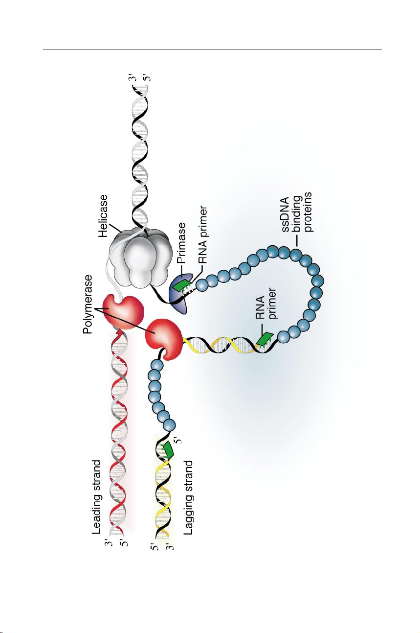
Figure 1 Role of DNA primase in semi-discontinuous DNA replication. A DNA replica-
tion fork is shown progressing from left to right to copy duplex DNA (black /gray). Because
the two strands of DNA are oriented in opposite polarities, only the leading strand (red ) is
synthesized continuously while the lagging strand ( yellow) is synthesized discontinuously.
If the two required DNA polymerases maintain contact to coordinate their activities, a loop-
ing of the lagging strand occurs. The DNA helicase (gray) separates the helix into ssDNA,
which is stabilized by ssDNA binding proteins (blue). The primase ( purple) synthesizes
RNA primers (green) on the lagging strand that are transferred periodically to the DNA polymerase (red ). P1: FNZ May 22, 2001 21:59 Annual Reviews ar131-02 DNA PRIMASES 41
by New York Medical College on 02/25/10. For personal use only.
Annu. Rev. Biochem. 2001.70:39-80. Downloaded from arjournals.annualreviews.org P1: FNZ May 22, 2001 21:59 Annual Reviews ar131-02 42 FRICK ! RICHARDSON
that DNA replication depends on a novel class of RNA-synthesizing enzymes
distinct from the classical RNA polymerases. Early studies of T7 DNA replication
implicated the phage gene 4 protein in priming DNA synthesis both in vivo (4, 5)
and in vitro (6, 7). Initially called a priming protein (8), the T7 gene 4 protein
later was designated a primase (9). The name has since been used to designate
its Escherichia coli counterpart, the DnaG protein, and all other members of this
enzyme class (10). Details of the role of DNA primase in the replication of the
lagging strand are presented in Figure 1.
This review focuses on the structure and function of DNA primases and on their
role in the initiation of Okazaki fragments. The coordination of primase activity
with the numerous proteins and enzymes that compose the replication complex
are also discussed. Three components of the replication complex in particular
greatly influence primase activity: DNA polymerase, DNA helicase, and ssDNA
binding protein. The importance of the interaction of primases with these proteins
is highlighted by the fact that in virtually all systems, one or more of these proteins
is physically associated with DNA primase or is essential for efficient primer
synthesis. Besides permitting semidiscontinuous replication, primases play other
significant roles in DNA synthesis and other cellular processes. For example,
primases initiate leading strand synthesis at chromosomal origins, and they are
involved in DNA repair (11). It should also be noted that DNA primases are not
the sole means of providing primers for DNA polymerases. Other mechanisms
for strand initiation include the use of DNA ends generated by recombination or
repair, transcripts made by conventional RNA polymerases, or priming proteins
attached to the ends of linear DNAs (12).
BASIC PROPERTIES OF PRIMASE GENES AND PROTEINS
With the recent explosion in genome sequencing, numerous proteins have been un-
covered that possess significant similarity to known primases. Most primases can
by New York Medical College on 02/25/10. For personal use only.
be classified into one of two large groups. One group contains the primases from
bacteria and their phages, and the second comprises eukaryotic DNA primases.
Annu. Rev. Biochem. 2001.70:39-80. Downloaded from arjournals.annualreviews.org
All primases share many properties, but the proteins in the two classes differ both
in structure and in their relationship with other proteins in the replication complex.
The prokaryotic primases are normally found associated with the replicative DNA
helicase, whereas the eukaryotic primases are found as complexes with DNA poly-
merase α (Pol α). Several examples of each class of primases as well as several
primases that do not fit into either of these two classes are discussed below. The
basic properties of primases are summarized in Table 1.
Prokaryotic Primases (Helicase Associated)
As shown in Figure 2, the prokaryotic primases include proteins from both bacteria
and their phages. The bacterial primases (Figure 2A) and the phage primases P1: FNZ May 22, 2001 21:59 Annual Reviews ar131-02 DNA PRIMASES 43
TABLE 1 Biochemical properties of purified DNA primases Amino Mr Primer Primer Recognition Source Gene acids (approx) length sequence sitea Prokaryotic E. coli dnaG 581 65,572 10–12 pppAG(N)8–10 5"-CTG -3" T7 gene 4 566 62,655 4–5 pppAC(N)2–3 5"-GTC -3" SP6 primase 661 74,096 4–10 pppGC(N)2–8 5"-GCA -3" T4 gene 61 342 39,768 4–5 pppAC(N)2–3 5"-GTT -3" P4 α 777 84,912 2–5 pppAG(N)0–3 5"-CT-3" Plasmid ColE2 rep 308 34,851 2–3 ppAGA 5"-TCTG -3" Viral Human HSV UL52 1,058 114,341 10–12 pppGG(N) 8–10 5"-ACCCTCC CGA -3" Eukaryotic S. cerevisiae PRI1 409 47,609 8–10 ppp(A/G)(N)7–9 PRI2 528 62,263 Drosophila PRI1 438 50,167 8–15 ppp(A/G)(N)7–14 melanogaster PRI2 533 61,390 Mus musculus PRI1 417 49,295 9–11 ppp(A/G)(N)8–10 PRI2 505 58,409 Homo sapiens PRI1 420 49,902 11–14 ppp(A/G)(N)8–13 PRI2 509 58,778
aCryptic nucleotides not copied into the primer are underlined. Eukaryotic primases have not been shown to use specific recognition sites.
(Figure 2B) are distantly related but share many biochemical properties, such as
a close association with DNA helicase and several consensus signature sequences (13–15).
by New York Medical College on 02/25/10. For personal use only. T7 Primase
The protein to which primase activity was first ascribed is the prod-
uct of gene 4 of bacteriophage T7 of E. coli. Gene 4 of phage T7 is essential
Annu. Rev. Biochem. 2001.70:39-80. Downloaded from arjournals.annualreviews.org
for phage replication (16), and T7 mutants defective in gene 4 synthesize a small
amount of DNA that hybridizes to only one strand (4, 5). A sensitive assay for
primer synthesis by the gene 4 primase was developed using circular ssDNA tem-
plates and T7 DNA polymerase. In this assay, DNA synthesis is dependent on
the presence of nucleoside triphosphates (NTPs) (6, 7) and the T7 gene 4 pro-
tein (9, 17). The T7 gene 4 primase requires only ATP and CTP to synthesize the
primers attached to DNA, which are predominantly of the sequence pppACN1N2,
where N1 and N2 are normally C or A (9, 17). G and U can also be incorpo-
rated into the third and fourth positions but at a much lower rate (18). In vivo,
it is tetraribonucleotides that are predominantly attached to the 5" termini of the
Okazaki fragments (19), and their sequences are the same as those synthesized P1: FNZ May 22, 2001 21:59 Annual Reviews ar131-02 44 FRICK ! RICHARDSON
by New York Medical College on 02/25/10. For personal use only.
Annu. Rev. Biochem. 2001.70:39-80. Downloaded from arjournals.annualreviews.org
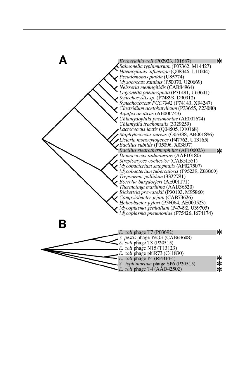
Figure 2 The prokaryotic DNA primases. (A) Bacterial encoding proteins significantly
similar to the E. coli DnaG primase. (B) Bacteriophages with genes significantly simi-
lar to the T7 63-kDa gene 4 DNA primase. Genetic database accession numbers for the
primase proteins are listed in parentheses. The amino acid sequences of putative DNA
primases were aligned to generate the phylogenetic trees depicted. Alignments of most of
the primases shown here are available on the World Wide Web courtesy of Mark Griep
(http://chem-mgriep2.unl.edu/replic/primase.html). Primases discussed in detail in this re- view are marked (∗). P1: FNZ May 22, 2001 21:59 Annual Reviews ar131-02 DNA PRIMASES 45
in vitro (20, 21). Each of the four deoxyribonucleotides is present at the RNA-
DNA junction of Okazaki fragments (17, 22, 23). Oligoribonucleotide synthesis
catalyzed by the T7 primase can also be measured directly by isolating the prod-
ucts synthesized in reactions not coupled to DNA synthesis (9). On large natural
ssDNAs, a mixture of di- (pppAC), tri- (pppACC/A), and tetraribonucleotides
(pppACCA/C and pppACAC/A) is synthesized. Similar products can also be
detected in reactions using oligonucleotide templates of defined length, provided
that appropriate template sequences, or primase recognition sites (see below), are present (24, 25).
In addition to its role as a DNA primase, the gene 4 protein is also a DNA heli-
case. T7 gene 4 actually encodes two colinear proteins: a full-length 63-kilodalton
(kDa) protein and a 56-kDa protein that is translated from an internal start codon
located 189 bases from the 5" end of the gene (26). The full-length 63-kDa gene
4 protein is both a primase and a helicase (27). The colinear 56-kDa gene 4 pro-
tein is a helicase but lacks primase activity (27). Only the 63-kDa gene 4 protein
is necessary and sufficient for T7 growth, although a mixture of the two forms
provides for the optimal rate of DNA synthesis in phage-infected cells (28, 29). E. coli Primase
Early studies on E. coli defective in the dnaG gene implicated
its product in the initiation of synthesis of Okazaki fragments both in vivo and
in vitro. The 60-kDa DnaG protein, initially purified using an in vitro comple-
mentation assay, was subsequently shown to catalyze the synthesis of small RNA
primers (30). To synthesize oligoribonucleotides efficiently on ssDNA, E. coli
primase requires either an origin coated with E. coli ssDNA binding protein or
the replicative helicase of E. coli (the product of the dnaB gene). Consequently,
the action of the E. coli primase has been studied in the context of two different
systems. The first system uses the G4 phage DNA (10), a close relative of φX174,
as a ssDNA template. On G4 ssDNA, synthesis is dependent on the presence of
E. coli ssDNA binding protein, and it begins at the site where replication of the
complementary strand begins in vivo, termed the G4 origin (G4oric) (31). The
by New York Medical College on 02/25/10. For personal use only.
RNA product is a 26- to 29-nucleotide-long transcript of G4oric (32). In the sec-
ond system, the activity of the DnaG protein is measured in the presence of the
E. coli DnaB helicase. This system has been designated the “general priming
Annu. Rev. Biochem. 2001.70:39-80. Downloaded from arjournals.annualreviews.org
reaction” because DnaB permits the primase to synthesize RNA primers on most
ssDNAs (33). In general priming reactions, the dnaG protein catalyzes the syn-
thesis of oligoribonucleotides 10 to 60 nucleotides long (34, 35), although most
are 11 residues (35). In both systems, primers begin with a pppAG dinucleotide
at the 5" end. Genes encoding DnaG-like DNA primases have also been identified
in many other organisms (Figure 2A).
Bacteriophage P4 Primase
Bacteriophage P4 is a temperate phage of E. coli that
can be maintained autonomously in the cell as a multicopy plasmid, and it encodes
some of its own replication apparatus (36). Mutations in the bacteriophage P4 α P1: FNZ May 22, 2001 21:59 Annual Reviews ar131-02 46 FRICK ! RICHARDSON
gene product do not support plasmid DNA synthesis (37). Characterization of a
purified P4 α protein revealed that the 88-kDa protein contains RNA polymerase
activity (38, 39). To synthesize primers, P4 α protein requires a DNA template,
NTPs, and Mg2+ and K+ ions for optimal activity (40–42). On ssDNA, the P4 α
protein synthesizes two- to five-nucleotide-long primers that begin with pppAG
(41). The P4 α protein, like the T7 gene 4 protein, is also a helicase. However,
unlike the T7 helicase, which translocates in a 5" to 3" direction, the P4 α protein
moves with a 3" to 5" directionality (41). P4 α protein possesses a third activity.
The protein binds duplex DNA with specificity for the six repeats of the sequence
5"-TGTTCACC-3" that are found at the P4 origin of replication (41). The primase,
helicase, and origin binding activities reside in three separate functional domains
mapping to the N terminus, a middle region, and the C terminus of the polypeptide, respectively (43, 44).
Bacteriophage T4 Primase
The requirement for NTPs in a T4 DNA replica-
tion system in vitro implicated RNA synthesis in the priming of DNA synthesis
(45). Dissection of these reconstituted systems revealed that primer synthesis was
dependent on the product of T4 gene 61 (gp61). T4 phage defective in gene 61
have a reduced rate of DNA synthesis (46) and accumulate ssDNA (47, 48). Like
the other primases in this class, the gp61 primase requires a helicase for optimal
activity, namely the T4 gene 41 protein (gp41) (49). The gp61 and gp41 have
molecular masses of 44 kDa and 58 kDa, respectively. In the presence of gp41
helicase, T4 primase synthesizes mainly pentanucleotides that begin with pppAC.
Pentaribonucleotides are the predominant primer species at the 5" ends of Okazaki
fragments during T4 DNA replication (49–51), and the Okazaki fragments initiate
with pppACNNN (50, 52). Because T4 gp61 cannot prime T4 DNA synthesis in
the absence of gp41, the functional primase can be considered a complex between
the gene 41 and gene 61 proteins. The two-protein complex requires the presence
of Mg2+ and K+ ions and ssDNA for the synthesis of oligoribonucleotides.
by New York Medical College on 02/25/10. For personal use only. SP6 Primase
Another novel prokaryotic primase was purified from Salmonella
typhimurium bacteriophage SP6, a distant cousin of phage T7. Phage SP6 is mor-
Annu. Rev. Biochem. 2001.70:39-80. Downloaded from arjournals.annualreviews.org
phologically similar to the E. coli phage T7, yet the viruses are so distantly related
that their two proteins share only 22% identical amino acids. The SP6 primase re-
quires GTP and CTP to synthesize oligoribonucleotides on ssDNA templates that
begin primarily with the sequence pppGC. Sequence comparisons have suggested
that, like the phage polymerases from T7 and P4, the SP6 primase protein may also
contain a replicative helicase function, although none has yet been identified (53).
Eukaryotic Primases (Polymerase α Associated)
The eukaryotic primases (Figure 3) typically purify as a complex of four pro-
teins: the 180-kDa DNA polymerase α (Pol α), a 70-kDa protein called the poly-
merase B subunit, and the two primase subunits of approximately 49 and 58 kDa, P1: FNZ May 22, 2001 21:59 Annual Reviews ar131-02 DNA PRIMASES 47
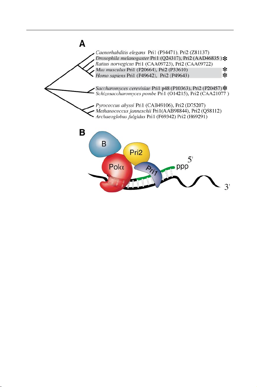
Figure 3 The eukaryotic family of DNA primases. (A) Organisms encoding proteins
similar to both the large and small subunits of human DNA primase. Cladograms were gen-
erated from alignments of Pri1 proteins. Alignments are available on Mark Griep’s web site
(http://chem-mgriep2.unl.edu/replic/EukPri1AA.html). Primases discussed in this review
are marked (∗). (B) Cartoon depicting the organization of the eukaryotic Pol α/primase
complex. The Pol α DNA polymerase (red ) and its B subunit (blue) form a complex with
the large primase subunit ( yellow), encoded by the Pri2 gene, and the small primase subunit
( purple), encoded by the Pri1 gene.
by New York Medical College on 02/25/10. For personal use only.
respectively. The smaller primase subunit contains the active site for oligoribonu-
cleotide synthesis. The genes encoding all of the subunits of the Pol α/primase
Annu. Rev. Biochem. 2001.70:39-80. Downloaded from arjournals.annualreviews.org
complex have been identified in humans, rats, mice, Drosophila, and the yeasts
Saccharomyces cerevisiae and Schizosaccharomyces pombe, although the primase
from S. pombe has not yet been purified or characterized. In addition, although
the genes are not yet cloned, the primase from calf thymus has been purified and
characterized. The recently sequenced genomes of several archaeons, particu-
larly those from the family Euryarchaeota, contain regions homologous to genes
encoding eukaryotic primases (Figure 3A). Yeast Primase
S. cerevisiae DNA primase copurifies with yeast DNA polymerase
I (Pol I), the Pol α equivalent in yeast (54, 55). A monoclonal antibody specific
for Pol I will coprecipitate this primase; the same antibody linked to a column P1: FNZ May 22, 2001 21:59 Annual Reviews ar131-02 48 FRICK ! RICHARDSON
can be used to separate the polymerase and primase subunits (55). Yeast primase
synthesizes primers that are 8–10 nucleotides long, and yeast Pol I extends these
primers at multiple sites on an M13 ssDNA template (56). Initiation begins only
on templates containing polypyrimidines (57), whereas the first NTP incorporated
at the 5" end of the primer is a purine (58). The complex is composed of 180-,
86-, 62-, and 48-kDa subunits; the two smaller subunits compose the primase.
The smallest, 48-kDa subunit (p48) alone catalyzes oligoribonucleotide synthesis.
Genetic analyses confirm that both primase subunits are essential for yeast survival
(59). The 62- and 48-kDa primase subunits physically interact with the 180-kDa
polymerase subunit but not with the 86-kDa B subunit, which binds only to the
180-kDa polymerase (60). The p48 and p58 subunits of the yeast primase are
encoded by two single-copy genes, PRI1 and PRI2, respectively. PRI1 is located on
chromosome IX (61) and PRI2 is located on chromosome XI (62, 63). Mutations
in either PRI1 or PRI2 result in severe defects in cell growth and DNA synthesis
that correlate with a high rate of mitotic recombination and spontaneous mutation
(64). Other yeast mutants have been selected that are lethal to strains carrying
primase defects. One of these mutants has been mapped to the MEC3 gene, which
is part of the G2 DNA damage checkpoint system, thus linking the primase with DNA repair (65).
Drosophila Primase
From Drosophila extracts, DNA Pol α purifies as a com-
plex with primase activity that has properties similar to the yeast primase discussed
above (66) and can likewise be purified using immunoaffinity chromatography
(67). The Drosophila Pol α/primase primes DNA synthesis on either poly(dT) or
M13 ssDNA templates (66) by synthesizing RNA primers that are 8–15 nucleotides
long (68). In the presence of deoxynucleoside triphosphates (dNTPs), Pol α
extends the primers at multiple nonrandom sites (66, 68). The Pol α/primase com-
plex is composed of four subunits of 182, 73, 61, and 50 kDa (69). The 182-kDa
subunit is the DNA polymerase, and the 61-kDa and 50-kDa subunits are required
for efficient primer synthesis (69). The genes encoding the DNA polymerase sub-
by New York Medical College on 02/25/10. For personal use only.
unit (70), its B subunit (71), and the large (72) and small (73) primase subunits
have been cloned and sequenced. The recombinant purified small (50-kDa) subunit
synthesizes oligoribonucleotides similar to those produced by the native complex
Annu. Rev. Biochem. 2001.70:39-80. Downloaded from arjournals.annualreviews.org
but is more thermally labile than the intact complex (73). Mouse Primase
Mouse primase was first purified using monoclonal antibodies
specific for human DNA Pol α (74, 75). The heterodimeric primase is part of a
complex containing the 180-kDa Pol α catalytic subunit and its 77-kDa B subunit
(76). The primase subunits are 58 kDa and 49 kDa and together synthesize oligori-
bonucleotides 9–10 nucleotides long that prime DNA synthesis (77). The mouse
primase requires NTPs and Mg2+ and synthesizes RNA on a variety of ssDNA
templates (78, 79). In the absence of DNA polymerase and dNTPs, mouse pri-
mase synthesizes multimeric oligoribonucleotides with a modal length of 9–10 nu-
cleotides (80). The genes for the primase subunits have been cloned and expressed P1: FNZ May 22, 2001 21:59 Annual Reviews ar131-02 DNA PRIMASES 49
in E. coli and baculovirus (81). The properties of the recombinant proteins are sim-
ilar to the native enzyme (82). The mouse primase 49-kDa and 58-kDa subunits
map to chromosome 10 in the distal part of band D, and to region A5-B of chro- mosome 1, respectively (83). Human Primase
Like the eukaryotic primases described above, the human
Pol α/primase (84, 85) can be purified from cell lysates using a monoclonal an-
tibody (86). In humans, both polymerase subunits are phosphorylated on serine
or threonine residues (87). The gene for the catalytic subunit of Pol α encodes
a 165-kDa protein that has sequence similarity with the E. coli Pol I family of
DNA polymerases (88). Due to its posttranslational modifications, human Pol α
migrates on polyacrylamide gels as a protein with a higher apparent Mr of 180–195
kDa. The other three subunits of the human Pol α/primase complex have masses
of 77, 59, and 50 kDa. The human primase complex synthesizes RNA primers
6–10 nucleotides long with ATP at the 5" end. When dNTPs are present in the
reaction, Pol α extends the primers to chains up to 3000 nucleotides long (86).
The cDNAs for all four subunits have been cloned and expressed to produce an
active recombinant complex (81, 89). The small primase subunit alone will syn-
thesize oligoribonucleotides but is very unstable. The activity of the small primase
protein can be stabilized by the addition of the large primase subunit or by the pres-
ence of divalent metal cations Mg2+ or Mn2+ during purification (89). The small
primase subunit maps to the long arm of chromosome 12 (90), and the large subunit
maps to two loci on chromosome 6 at bands 6p11.1–p12 (91). Miscellaneous Primases
Not all known primases are homologous to proteins in either of the two classes
outlined above. One set of these miscellaneous primases comprises viral proteins
that are immunologically distinct from the eukaryotic primases. The members
of another class are the products of plasmid sog genes. These “suppressors of
by New York Medical College on 02/25/10. For personal use only.
dnaG” permit the growth of strains carrying temperature-sensitive alleles of dnaG
at nonpermissive temperatures. Some, but not all, plasmid-encoded primases share
homology with prokaryotic primases, but they also contain unique properties that
Annu. Rev. Biochem. 2001.70:39-80. Downloaded from arjournals.annualreviews.org
distinguish them from other primases.
Transfer Protein Primases
The first plasmid-encoded primases described were
a series of proteins that are transported from donor to recipient E. coli cells during
conjugation and are encoded by plasmids from the IncI and IncP compatibility
groups (92). One of these transfer proteins is the product of the sog gene of the
IncI plasmid ColIb-P9. As is the case with T7 gene 4, the sog gene encodes two
polypeptides of 210 kDa (Sog210) and 160 kDa (Sog160) from separate in-frame
translational start sites (93). The primase function is part of the amino terminus
of the 210-kDa protein (93, 94). Two other transfer protein primases (called TraC
proteins) are encoded by plasmids from the IncP group. One is the TraC protein P1: FNZ May 22, 2001 21:59 Annual Reviews ar131-02 50 FRICK ! RICHARDSON
of plasmid RP4 (IncPα), and another is the TraC protein of plasmid R751 (IncPβ)
(95). Again, several forms of the TraC protein are encoded from different start sites.
The RP4 traC gene encodes the 116-kDa TraC1 protein and the 81-kDa TraC2 pro-
tein (96). Plasmid RP751 traC encodes four proteins: TraC1 (192 kDa), TraC2 (152
kDa), TraC3 (135 kDa), and TraC4 (83 kDa). Even the smallest, TraC4, is a func-
tional primase (95), which suggests that in TraC proteins the primase domain likely
resides in the C terminus. Segments of all these transfer proteins share sequences
with the primase domain of the P4 primase (93), but only these small regions are
similar to other prokaryotic primases. The other domains of these large proteins
likely facilitate the transfer of genetic material from one bacterium to another.
ColE2 Rep Primase
Another plasmid gene that encodes a novel primase is the
rep gene of plasmid ColE2 (97). At 35 kDa, the Rep primase is smaller and
lacks sequence homology with other primases. Remarkably, this small protein can
prime DNA synthesis in the presence of plasmid DNA, E. coli DNA polymerase I,
E. coli ssDNA binding protein, ATP, GTP, ADP, and dNTPs (98). The Rep pro-
tein primes DNA synthesis with ppAGA opposite the sequence 5"-TCTG-3" in
the plasmid origin. Synthesis requires ADP, ATP, and GTP, and products retain
[β-32P]ADP at their 5" end, suggesting that RNA synthesis begins opposite the
dT in the recognition site (99). Binding of the ColE2 Rep primase to the plasmid
origin is so specific that it does not bind to the origin of the related plasmid ColE3
(100), the two origins differing by only 2 of 33 base pairs (97). Two regions near
the C terminus of the Rep primase are likely involved in sequence-specific DNA
binding (101). Unlike other known primases, the Rep primase facilitates DNA
synthesis on a circular duplex DNA molecule.
Herpes Simplex Virus Primase
Evidence that herpes simplex virus (HSV) en-
codes its own DNA primase was first found in HSV-infected HeLa cells (102).
The HSV primase exists as a three-polypeptide complex consisting of the UL52,
UL5, and UL8 gene products (103) that also contains 5"-3" helicase activity (104).
by New York Medical College on 02/25/10. For personal use only.
A subassembly of the UL5 (97-kDa) and UL52 (120-kDa) proteins is sufficient
for helicase and primase activity (105, 106). The UL52 protein contains the cat-
alytic center for primase activity (107), and the UL5 subunit is likely the helicase
Annu. Rev. Biochem. 2001.70:39-80. Downloaded from arjournals.annualreviews.org
(108, 109). The herpes UL5/UL52 complex synthesizes 10- to 13-nucleotide-
long primers on M13 ssDNA and unwinds DNA at a rate of two base pairs per
second (110). In contrast with the host eukaryotic primase, HSV primase does
not reinitiate from these primers to make primer multimers (110). Although it
is not essential, the presence of the UL8 protein stimulates primer synthesis by
the UL5 and UL52 components (111, 112), especially on templates coated with
the herpes ssDNA binding protein, ICP8 (113). Similar genes have been iden-
tified in other viruses including equine HSV, human Epstein-Barr virus, human
cytomegalovirus, and varicella-zoster virus. Alignments of these viral primases
reveal amino acids likely to be critical for primer synthesis. One such align-
ment is available on the web site of Mark Griep at the University of Nebraska
(http://chem-mgriep2.unl.edu/replic/VirPrAA.html). P1: FNZ May 22, 2001 21:59 Annual Reviews ar131-02 DNA PRIMASES 51
INTERACTIONS BETWEEN PRIMASES AND DNA TEMPLATES
Primase must first bind a DNA template before synthesizing RNA primers (25, 114).
The eukaryotic Pol α/primase complexes bind DNA with a Kd of 0.1 to 1.0 µM
and protect 9 nucleotides of the primer and 13 nucleotides of the template from
nuclease digestion (114, 115). To enable frequent primer synthesis on the lagging
strand, most primases will synthesize oligoribonucleotides complementary to vir-
tually any ssDNA to which they are bound. Experiments using several prokaryotic
and viral primases have shown, however, that certain DNA sequences support a
dramatically higher rate of primer synthesis. These sequences, termed primase
recognition sites, differ for each of the primases examined and may play an im-
portant role in the coordination of lagging strand synthesis. Despite not having
a stringent requirement for a specific recognition site, many eukaryotic DNA pri-
mases likewise display sequence specificity.
Prokaryotic and Viral Primase Recognition Sites
With φX174 (22) or M13 ssDNA (23) as a template, T7 primase enables T7 DNA
polymerase to initiate DNA synthesis primarily at 13 and 9 sites, respectively.
Most of the T7 primase recognition sites contain the sequences 5"-GGGTC-3",
5"-TGGTC-3", or 5"-GTGTC-3", which direct the synthesis of the primers
pppACCC, pppACCA, and pppACAC, respectively. The identities of the tem-
plate recognition sites have been confirmed in vivo and in vitro by mapping the
exact locations of five primer RNA to DNA transition sites on a fragment from
the T7 genome (116) and using short synthetic oligonucleotide templates (24, 25).
All T7 primase recognition sites contain the same trinucleotide, 5"-GTC-3". Primer
synthesis begins opposite the 3" dT, and the 3" cytosine is cryptic, meaning that its
complement is not incorporated into the oligoribonucleotide products. No RNA
synthesis is supported by short synthetic templates lacking either the cryptic dC
by New York Medical College on 02/25/10. For personal use only.
or the adjacent dT in the recognition site (25). Templates containing modified dC
or dT bases likewise support a lower rate of oligoribonucleotide synthesis than
sequences containing the preferred recognition site 5"-GTC-3" (117, 118). In par-
Annu. Rev. Biochem. 2001.70:39-80. Downloaded from arjournals.annualreviews.org
ticular, templates lacking the N3 nitrogen of the cryptic dC or the N3 nitrogen of
dT support little detectable synthesis, highlighting potential interactions between
the primase and functional groups involved in Watson-Crick base-pair formation (118).
Other phage primases preferentially initiate synthesis at different characteristic
primase recognition sites. The SP6 primase begins primers with pppGC preferen-
tially at sites containing the sequence 5"-GCA-3" (53). The T4 primase recognizes
the site 5"-GTT-3" or 5"-GCT-3" depending on certain conditions. On M13 ssDNA,
all initiation sites share the sequence 5"-GTT-3" or 5"-GCT-3" and give rise to the
primers pppACNNN and pppGCNNN (119). Only the 5"-GTT-3" site is used on
T4 DNA that contains hydroxymethylcytosine (120). The first dT in the recog-
nition site is cryptic. Synthesis by the P4 primase begins at the phage origin of P1: FNZ May 22, 2001 21:59 Annual Reviews ar131-02 52 FRICK ! RICHARDSON
replication, where the P4 α typically starts primers with pppAG at the two-base sequence 5"-CT-3" (41).
The bacterial E. coli DnaG primase also recognizes a trinucleotide sequence in
DNA. On the G4oric template, the E. coli DnaG protein catalyzes the synthesis of
26- to 29-nucleotide-long oligoribonucleotides (121, 122) that begin with pppAG
at the sequence 5"-CTG-3" (123). G4oric consists of three stem-loop structures that
lie on the 5" side of the trinucleotide 5"-CTG-3" sequence and direct the binding of
E. coli ssDNA binding protein to the DNA. None of the three hairpins normally
present in G4oric are required for DnaG protein to initiate primer synthesis, but
the sequence 5"-CTG-3" is required (124). Other synthetic ssDNA templates con-
taining 5"-CTG-3" but not any of the remaining G4oric sequence likewise support
DnaG-catalyzed primer synthesis (124). This trinucleotide sequence is also rec-
ognized by DnaG protein at the origins of related phages St-1, φK, and α3 (125).
Although early models invoked secondary structures of G4oric as the recognition
site for the primase, later studies strongly support a model in which the hairpin
loops help expose the primase recognition site in the presence of E. coli ssDNA
binding protein (126). The E. coli primase recognition site 5"-CTG-3" remains
free of ssDNA binding protein (126) and can interact directly with the primase
(127). A second primase molecule binds to a region flanking the 5" end of the
primase recognition site, which also remains free of ssDNA binding protein (128).
Thus, although DnaG protein is a monomer in solution (30, 129), a dimer may be
required to support replication at replication origins (130).
Clearly, prokaryotic primases preferentially synthesize primers at certain DNA
sequences. However, different sequences are recognized by the various proteins:
5"-CTG-3" by the E. coli DnaG protein, 5"-GTT-3" (or 5"-GCT-3") by the T4 gene
61 protein, 5"-GCA-3" by the SP6 primase, and 5"-GTC-3" by the T7 primase. In
each case, a trinucleotide sequence, in which the 3" residue is cryptic, is required
for recognition. The biological role of these trinucleotide primase recognition sites remains unclear.
Although it is not evolutionarily related to the prokaryotic primases, the herpes
by New York Medical College on 02/25/10. For personal use only.
simplex primase has also been shown to synthesize primers preferentially at spe-
cific sequences (112). On φX174 DNA, purified herpes primase predominantly
synthesizes primers of the sequence 5"-GGGAGGGUAG-3" beginning opposite
Annu. Rev. Biochem. 2001.70:39-80. Downloaded from arjournals.annualreviews.org
the 3" dC in the template sequence 5"-ACCCTCCCGA-3". Only oligonucleotides
containing this recognition sequence will inhibit RNA primer DNA synthesis by
the HSV primase/polymerase complex (131).
Sequence-Specific Interactions by Eukaryotic Primases
Eukaryotic primases require the presence of pyrimidines in the ssDNA template for
activity and bind polypyrimidine templates more tightly than they do polypurine
templates (132, 133). This common characteristic is likely due to the fact that all
primers are initiated with purine NTPs (134). However, on templates containing a
mixture of purines and pyrimidines, eukaryotic primases do not initiate synthesis P1: FNZ May 22, 2001 21:59 Annual Reviews ar131-02 DNA PRIMASES 53
randomly but prefer certain sites. The most convincing evidence of eukaryotic
primase sequence specificity comes from experiments that mapped specific pri-
mase initiation sites on simian virus 40 (SV40) DNA to a region located near the
65-base minimal origin of replication (79, 134–137). On SV40 DNA, replication
begins almost exclusively at template dTs and is spaced regularly within, and to
the 3" side of, the 65-base-pair minimal origin (134–137). When given a ssDNA
template made from the strand encoding the SV40 early mRNA, mouse primase
begins synthesis at only five sites, four of which are clustered within 10 nucleotides
of each other. On the other strand encoding the late mRNA, primase synthesizes
primers at six sites adjacent to the 65-nucleotide minimal origin (79). Deletion of
only six nucleotides in this origin significantly affects initiation site usage (137).
Mouse primase initiation has also been studied using ssDNA templates de-
rived from the minute virus of mice. On these DNAs, primer synthesis begins
mainly at 17 sites that all share a template sequence (5"-CCA-3" or 5"-CCC-3")
2–14 nucleotides downstream from the start site for primer synthesis (138, 139).
Mouse primase binds DNA with these trinucleotide sequences more tightly than it
does DNA lacking these sequences and has the highest affinity for templates con-
taining 5"-CCC-3" approximately 10 nucleotides downstream from the initiation
sites for primer synthesis (139). Similar pyrimidine trinucleotides have also been
identified in initiation sites used by primases purified from simian cells (134) and
HeLa (140) cells. Thus, like the prokaryotic primases, human and mouse primases
recognize a trinucleotide. However, whereas prokaryotic primases initiate synthe-
sis directly opposite to their recognition sequences, eukaryotic primases initiate
primers opposite to nucleotides flanking the 3" end of the recognition sequence.
In a study using a Pol α/complex isolated from calf thymus, Suzuki et al (141)
found that only 9 out of 140 ssDNA templates screened support detectable primer
synthesis. The active templates contained an unusually high percentage of pyrim-
idines. Substitutions of adenylates for the pyrimidines in the active templates affect
both the rate of primer synthesis and the affinity of the primase for the template.
A similar template specificity was observed using primase free of Pol α (142).
by New York Medical College on 02/25/10. For personal use only.
In these studies, the initiation of primer synthesis seems to require a six-base
pyrimidine-rich tract of DNA, bounded by a 3" purine, but unlike the mouse pri-
mase initiation sites, the calf thymus sites share no commonly conserved sequence
Annu. Rev. Biochem. 2001.70:39-80. Downloaded from arjournals.annualreviews.org
(141). The nonrandom primer initiation by calf thymus Pol α/primase has been
confirmed using other, longer templates (143). Initiation site usage is dependent
on the reaction conditions and appears more random at higher concentrations of
NTPs (144) and when Mn2+ is added to the reaction (145). PRIMER SYNTHESIS
Primases catalyze the synthesis of oligoribonucleotides in a minimum of five
discrete steps: template binding, NTP binding, initiation, extension to a functional
primer, and primer transfer to DNA polymerase. We have proposed a simple P1: FNZ May 22, 2001 21:59 Annual Reviews ar131-02 54 FRICK ! RICHARDSON
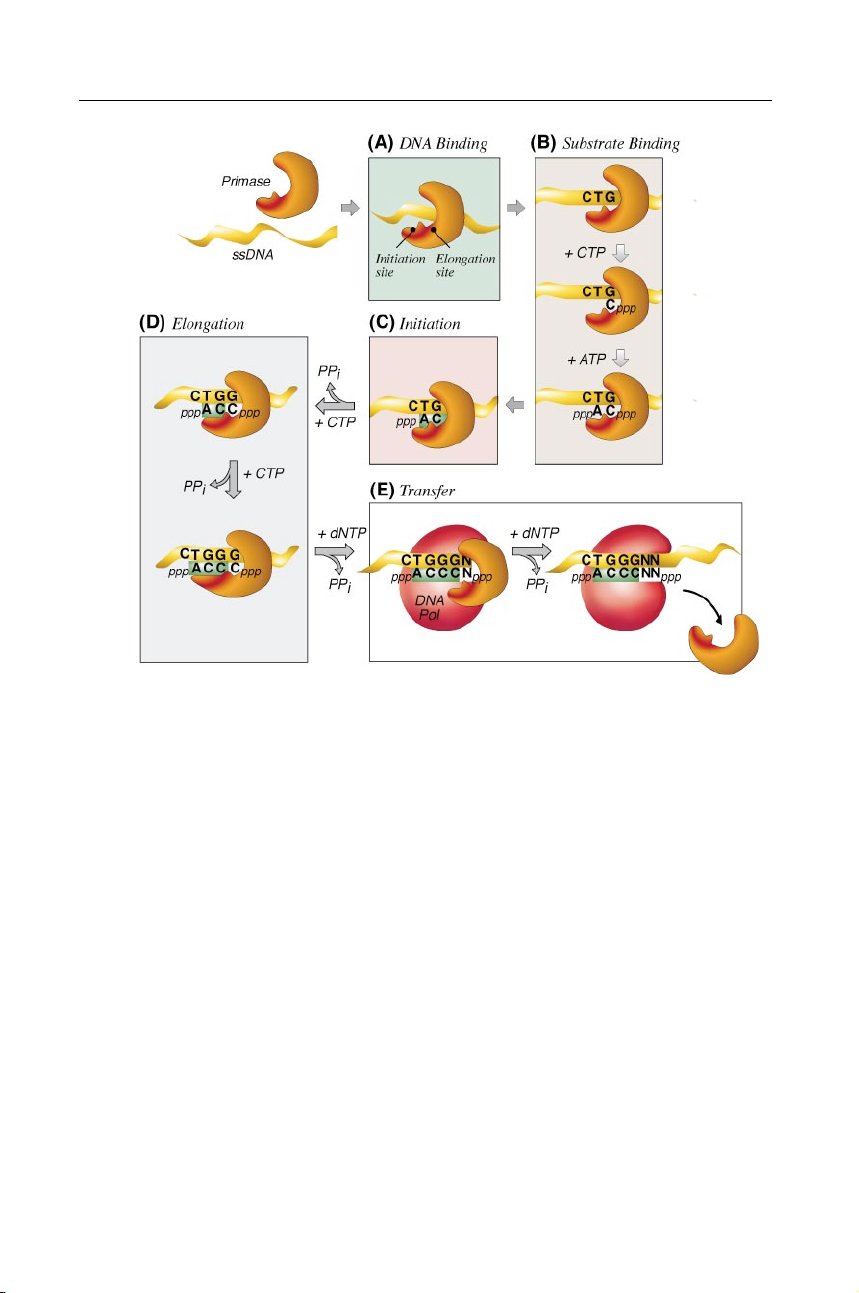
Figure 4 Steps in primer synthesis. This hypothetical mechanism is intended to depict
primer synthesis by the T7 primase. Other primase mechanisms differ concerning NTPs
used, primer sequence, primer length, and template recognition site. (A) DNA primase
(orange) binds to ssDNA ( yellow). (B) When the primase encounters an appropriate initi-
ation site, two NTPs bind. The first NTP binds to the elongation site, eventually becoming
the second nucleotide in the primer. The second NTP binds to the initiation site, and is
incorporated at the 5" end of the primer. (C) Primer synthesis is initiated by the formation
by New York Medical College on 02/25/10. For personal use only.
of a dinucleotide and inorganic pyrophosphate from the two bound NTPs. (D) The growing
oligoribonucleotide is transferred to the initiation site while additional NTPs bind to the
elongation site; nucleotides are incorporated at the 3" end of the primer. (E ) Primer RNA
Annu. Rev. Biochem. 2001.70:39-80. Downloaded from arjournals.annualreviews.org
is transferred to the replicative DNA polymerase that adds deoxynucleotides derived from dNTPs to their 3" ends.
mechanism that involves two NTP binding sites on the primase protein (146).
In this mechanism (Figure 4), the site at which the NTP is to be incorpora-
ted at the 5" end of the primer is referred to as the initiation site. The second
site, which binds the NTP added to the 3" end of the primer, is referred to as the
elongation site. At each elongation step of primer synthesis, the product (n + 1)
oligonucleotide must be transferred to the initiation site so that another NTP may
bind to the elongation site. The length of the oligonucleotide that can bind in the P1: FNZ May 22, 2001 21:59 Annual Reviews ar131-02 DNA PRIMASES 55
initiation site may limit the final length of the oligoribonucleotide synthesized by the primase.
Substrate Binding and Primer Initiation
After binding a DNA template (Figure 4A), primase must next bind two NTP
substrates (Figure 4B) to catalyze the formation of a dinucleotide and inorganic
pyrophosphate (Figure 4C). The NTP incorporated at the 5" end of the primer is
usually a purine, ATP or GTP, and retains its 5" triphosphate moiety after dinu-
cleotide synthesis. Most primases show little preference regarding the triphosphate
end of the NTP incorporated at the 5" end of the primer. NTP analogs with modi-
fied 5" phosphate groups (146–148) and even nucleotides cross-linked to proteins
via their 5" phosphates are incorporated at the 5" end of primers (149, 150).
In the phage T7 system, primers usually begin with pppAC. However, in the
absence of DNA, T7 primase synthesizes other dinucleotides at a low rate in a re-
action that is enhanced greatly when Mn2+ is present (151, 152). In these template-
independent reactions, CMP predominates at the 3" end of the dinucleotide, but
each of the four canonical NTPs is incorporated at the 5" end, suggesting that
during initiation, CTP binds first and helps to locate primase recognition sites in
DNA (153). Indeed, the affinity of T7 primase for DNA templates increases when
primase is bound to either ATP or CTP (25). The Km for CTP in the reaction
catalyzed by T7 primase (0.8 mM) is higher than the cellular concentration of
CTP, suggesting that the concentration of CTP could contribute to control of the
rate of primer synthesis (146). Under optimal conditions, T7 primase can initiate
primers at a rate of four dinucleotides per second (25), a rate sufficient to facilitate T7 DNA synthesis (154).
The kinetics of primer synthesis catalyzed by E. coli primase have been exam-
ined using a 23-nucleotide DNA template, which contains the primase recognition
site 5"-GTC-3, the minimal template required for full-length (10- to 12-nucleotide)
primers (155). In this system, under steady-state conditions, mainly full-length
by New York Medical College on 02/25/10. For personal use only.
products are synthesized, and intermediates do not accumulate, suggesting that
the rate-limiting step occurs before the synthesis of the dinucleotide. The rate of
primer synthesis by E. coli primase on a 23-nucleotide template is quite slow at
Annu. Rev. Biochem. 2001.70:39-80. Downloaded from arjournals.annualreviews.org
0.00089 sec−1 (155). However, when the E. coli DnaB helicase is present, the rate
of primer synthesis increases 15-fold (156).
Reported Km values for NTPs in reactions catalyzed by eukaryotic primase
range from 8 to 175 µM (78, 115, 133, 157, 158). The rate-limiting step during the
synthesis of eukaryotic primers occurs before or during dinucleotide synthesis.
With the calf thymus primase, after the formation of an E • DNA • NTP • NTP
complex, dinucleotides are synthesized at a rate of 0.003 s−1 and are then rapidly
extended to full-length primers (159).
As shown in Figure 4B, the NTPs may bind the primase in a particular order,
with the first NTP binding to the primase at the elongation site. The best evidence P1: FNZ May 22, 2001 21:59 Annual Reviews ar131-02 56 FRICK ! RICHARDSON
supporting this binding order comes from studies using eukaryotic primases.
Because eukaryotic primases do not initiate solely at specific primase recogni-
tion sites, they may incorporate either ATP or GTP at the 5" end of the primer.
By identifying the 5"-terminal nucleotide in primers synthesized at various NTP
concentrations, Sheaff & Kuchta have demonstrated that the first NTP that binds
calf thymus primase becomes the second nucleotide in the primer and determines
where on the template synthesis begins (159). Primer Elongation
During strand elongation, the growing oligoribonucleotide is transferred to the
initiation NTP binding site to make room for another NTP to bind to the elon-
gation site and subsequently attach to the 3" hydroxyl of the oligoribonucleotide
(Figure 4D). The length of oligoribonucleotides that are synthesized varies among
the different primases and conceivably could be regulated by the length of the
oligoribonucleotide that can be accommodated at the initiation NTP binding site
of the enzyme. Oligoribonucleotides normally used as primers by DNA poly-
merase are designated full-length or unit-length primers. Unit-length primers
are synthesized processively (80, 115, 146, 160) but are often not the only prod-
ucts observed in vitro. Shorter “abortive primers” accumulate with all primases,
and under certain conditions, eukaryotic primases synthesize primer multimers
by adding nucleotides to the ends of unit-length primers. Primer multimers are
normally the length of multiple unit-length primers. For example, if the primase
synthesizes primers 10 nucleotides long, then primer multimers would be 20, 30, or 40 nucleotides long.
The size of unit-length primers varies depending on each primase and the needs
of its cognate replicative polymerase. Phage primases synthesize the shortest pri-
mers, normally fewer than 5 nucleotides. Most other primases synthesize primers
slightly longer than 10 nucleotides (Table 1). During steady-state RNA synthesis
catalyzed by primase, the quantities of the various products do not change over
by New York Medical College on 02/25/10. For personal use only.
time, indicating a processive reaction (115, 146, 160, 161). Significant amounts
of nucleotides are incorporated into abortive products, but the relative amount
of shorter products can be minimized in conditions favoring the addition of the
Annu. Rev. Biochem. 2001.70:39-80. Downloaded from arjournals.annualreviews.org
next nucleotide (146, 160). The physiological significance of abortive products
remains elusive, but primases are able to rebind and elongate oligonucleotides,
suggesting that abortive primers could serve as intermediates in primer synthesis (147).
Primer multimers are synthesized in reactions containing purified eukaryotic
primases free of associated polymerase activity, or in reactions containing the Pol
α/primase complex but free of dNTP substrates. For example, in the absence of
Pol α, mouse primase continues to synthesize primers in modal increments of
the original 9- to 11-base RNA primer up to 50 bases in length (78, 80). Yeast
primase likewise rebinds and extends unit-length primers to synthesize multimeric
primers during repeated cycles of processive oligoribonucleotide synthesis (161). P1: FNZ May 22, 2001 21:59 Annual Reviews ar131-02 DNA PRIMASES 57
Multimeric primers are synthesized more frequently at low temperatures, which
would favor the re-annealing of unit-length primers (160).
Several DNA primases select and incorporate NTPs with low fidelity. The
E. coli primase can incorporate deoxynucleotides as well as ribonucleotides into
primers and will add ribonucleotides to the 3" hydroxyl of a DNA chain (34, 156,
162–164). The biological significance of these in vitro observations is not known,
nor is it known if such mixed primers occur in vivo. Because of its ability to
incorporate deoxyribonucleotides into primers, the DnaG protein can also incor-
porate dideoxyribonucleotides into primer chains, thus preventing their further
extension (164). Besides lacking the ability to discriminate between NTPs and
dNTPs, E. coli primase also lacks fidelity in the selection of the appropriate nu-
cleotide base (128, 165). E. coli primase forms non–Watson-Crick base pairs with
a rate approaching 1 in every 10 nucleotides polymerized (155). The E. coli pri-
mase shares low fidelity with eukaryotic primases, notably Drosophila primase,
which incorporates deoxynucleotides and frequently synthesizes primers not com-
plementary to the template (166). Low fidelity is not, however, a universal feature
of all primases. For example, T7 primase differentiates between ribo and deoxy
sugars and will misincorporate nucleotides at a detectable rate only when solely
the incorrect nucleotide is present in a reaction (146).
Transfer of Primers to DNA Polymerase
To fulfill their primary function, DNA primases must not only initiate and elongate
oligoribonucleotides but also transfer primers to DNA polymerase (Figure 4E ).
The communication between primase and DNA polymerase is most apparent when
primer synthesis is examined in the presence and absence of DNA polymerase.
In eukaryotic systems, where the primase and polymerase form a tight complex,
the two activities are clearly interconnected. In the presence of ssDNA template,
NTPs, and dNTPs, eukaryotic primase transfers primers directly to DNA Pol α
without dissociating from the template or from the enzyme complex (167).
by New York Medical College on 02/25/10. For personal use only.
Whereas in eukaryotic systems primases and polymerases form tight complexes,
in the prokaryotic replication fork, DNA polymerases and primases are more
transiently related. The primase repeatedly associates and dissociates from the
Annu. Rev. Biochem. 2001.70:39-80. Downloaded from arjournals.annualreviews.org
replication fork while catalyzing multiple rounds of primer synthesis (168–170).
In E. coli, the DNA polymerase III (Pol III) enzyme complex influences primer
synthesis, probably by competing with the primase for the primer 3" end. In the
presence of Pol III, the most abundant product is 8 nucleotides long instead
of 11, and longer oligoribonucleotides are not synthesized (35, 162). In a com-
plete system containing primase, Pol III, and dNTPs, the primers at the ends of
Okazaki fragments have been found to be 1 to 9 nucleotides long in one study
(171) and 9 to 14 in another (35). Yuzhakov et al (172) have proposed an ele-
gant mechanism involving a three-point switch to explain the transfer of the
primer from the E. coli primase to Pol III during DNA replication. They have
demonstrated that Pol III, with its sliding-clamp processivity subunit β, cannot P1: FNZ May 22, 2001 21:59 Annual Reviews ar131-02 58 FRICK ! RICHARDSON
assemble onto primed DNA while the primase is still bound. To load onto a
primed template, the Pol III χ subunit, part of the clamp-loading γ complex,
makes contact with ssDNA binding protein, which coats the template. Because
DnaG primase also contacts ssDNA binding protein to assemble properly on DNA,
Yuzhakov et al have proposed that the χ subunit may act to displace the primase
from the replication fork. Thus, ssDNA binding protein facilitates primer transfer
by switching its contact between primase and the Pol III χ subunit (172). Inhibitors
Inhibitors of DNA primases are valuable compounds not only for the elucida-
tion of biochemical pathways but also as lead compounds to design drugs that
block proliferative, bacterial, or viral diseases. Most of the compounds known
to inhibit primases are nucleotide analogs. For example, E. coli primase will use
2",3"-dideoxynucleoside 5"-triphosphates (ddNTPs) as substrates, and when in-
corporated, these compounds act as chain terminators (124). Another nucleotide
analog, 2"-deoxy-2"-azidocytidine, potently inhibits DnaG and is incorporated into
the primer, thus severely limiting primer extension (173). The T7 primase will not
use either dNTPs or ddNTPs, but it will incorporate 3"-dCMP as a chain terminator,
albeit at a rate 1/100 that of CMP (146).
The triphosphate derivatives of the anticancer drug cytarabine (cytosine arabi-
noside, araC) and the antiviral drug vidarabine (adenosine arabinoside, araA) both
inhibit DNA primases (133, 158, 174, 175). AraATP is a potent inhibitor of calf
thymus DNA primase with a Ki of approximately 2 µM (158) and is preferred
by the enzyme over ATP, 2"-dATP, 3"-dATP, or ddATP (174). With the human eu-
karyotic primases, araCTP (1-β-D-arabinofuranosylcytosine 5"-triphosphate) and
araATP (9-β-D-arabinofuranosyladenosine 5"-triphosphate) compete with GTP,
with Ki values of 122 and 128 µM, respectively (133).
The potency of araCTP and araATP in inhibiting human primase is surpassed
by halogenated nucleotide analogs, including FaraATP (9-β-D-arabinofuranosyl-
by New York Medical College on 02/25/10. For personal use only.
2-fluoroadenosine 5"-triphosphate), and the even more potent inhibitors FMAC-TP
[1-(2-deoxy-2-fluoro-β-D-arabinofuranosyl)-5-methylcytosine 5"-triphosphate],
FIAC-TP [1-(2-deoxy-2-fluoro-β-D-arabinofuranosyl)thymine 5"-triphosphate],
Annu. Rev. Biochem. 2001.70:39-80. Downloaded from arjournals.annualreviews.org
and 2"-F-araTTP [1-(2-deoxy-2-fluoro-β-D-arabinofuranosyl)thymine 5"-triphos-
phate] (133). FaraATP is a noncompetitive inhibitor of primer synthesis with a Ki
25-fold lower than the Km of ATP (176). Eukaryotic primases will preferentially
incorporate FaraATP into RNA primers 30-fold more efficiently than they do ATP.
Once incorporated, FaraATP acts as a chain terminator. As a result, in the presence
of FaraATP, large amounts of short abortive oligoribonucleotide are produced that
are not extended to full-length primers (177). In addition, (dichloroanilino)purines
and -pyrimidines selected to inhibit the herpes simplex virus type 1 helicase-
primase have been shown to slow the replication of HSV1 in infected HeLa cells (178).