Biology Lab Manual - January 2024 Edition | Môn Biology - Trường Đại học Quốc tế, Đại học Quốc gia Thành phố Hồ Chí Minh
Before doing lab-work, students should be aware of the risks, hazards and safety conditions maintained in laboratory. Tài liệu được sưu tầm gồm 38 trang, giúp bạn ôn tập tốt hơn. Mời các bạn đón xem.
Môn: Biology 10 tài liệu
Trường: Trường Đại học Quốc tế, Đại học Quốc gia Thành phố Hồ Chí Minh 2 K tài liệu
Tác giả:


Preview text:
lOMoAR cPSD| 59078336 SCHOOL OF BIOTECHNOLOGY January 2024 version. BIOLOGY (BT312IU) Labwork Manual
[ G R O U P N U M B E R ] _ _ _ _ _ _ _ _ _ _ _ _ _ _ _ _ _ _ _ _ _ _ _ _ _ _ _
[ G R O U P M E M B E R S ]
_ _ _ _ _ _ _ _ _ _ _ _ _ _ _ _ _ _ _ _ _ _ _ _ _ _ _ _ _ _ _ _ _ _ _ _
_ _ _ _ _ _ _ _ _ _ _ _ _ _ _ _ _ _ _ _ _ _ _ _ _ _ _ _ _ _ _ _ _ _ _ _
_ _ _ _ _ _ _ _ _ _ _ _ _ _ _ _ _ _ _ _ _ _ _ _ _ _ _ _ _ _ _ _ _ _ _ _
_ _ _ _ _ _ _ _ _ _ _ _ _ _ _ _ _ _ _ _ _ _ _ _ _ _ _ _ _ _ _ _ _ _ _ _ 0 1 lOMoAR cPSD| 59078336 GENERAL ISSUES GRADING Labwork assessments Prelab (individually) 30% Lab report (in group) 40%
Lab examination (individually) 30% Total 100% CODE OF CONDUCT
1. Students must understand, remember and follow the Laboratory safety (see next page) before starting this course.
2. Absence without permission is not allowed, due to the nature of the labs. For make-up class, join
another group having that practical session.
3. Pre-labs must be submitted at 8:15AM or 1:15PM on each practical class.
4. Examination is composed of practical performance and questions answering.
*** More information, please go to: OUTLINES LABORATORY SAFETY PRACTICAL 1:
MICROSCOPY AND CELL OBSERVATION PRACTICAL 2:
ORGANIC COMPOSITIONS OF THE CELL PRACTICAL 3:
PHOTOSYNTHESIS AND TRANSPIRATION PRACTICAL 4: OSMOSIS AND LEAF STOMATA PRACTICAL 5: ENZYMES PRACTICAL 6:
CELL DIVISIONS-MITOSIS & MEIOSIS FINAL EXAM: WRITING AND PRACTICAL SKILL GUIDELINE FOR LAB REPORT LABORATORY SAFETY
Before doing lab-work, students should be aware of the risks, hazards and safety conditions
maintained in laboratory. Risk is identified as a substance or biological agent that might be harmful
under specific circumstances while hazard is the ability of a substance or biological agent to cause harm. lOMoAR cPSD| 59078336
In any case, a person working with chemicals, bio-chemicals or other related agents should follow strictly
certain principles applied in each laboratory, and all practical works must be carriedout with safety in
mind to minimize the risk of harm to yourself and to others. The followings are the basic rules for laboratory work. 1.
Make sure that you know what to do in case of fire, including exit routes, how to raise
the alarm, and where to gather after leaving the building. Remember that the most important
consideration at all time is human safety. 2.
Follow tutor’s instructions and the laboratory principles while doing lab work. 3.
Wear lab-coat and closed footwear at all time. 4.
Do not smoke, eat or drink in laboratory because of the risks of contamination by inhalation or ingestion. 5.
Do not mouth-pipette any liquid. 6.
Take care when handling glassware. 7.
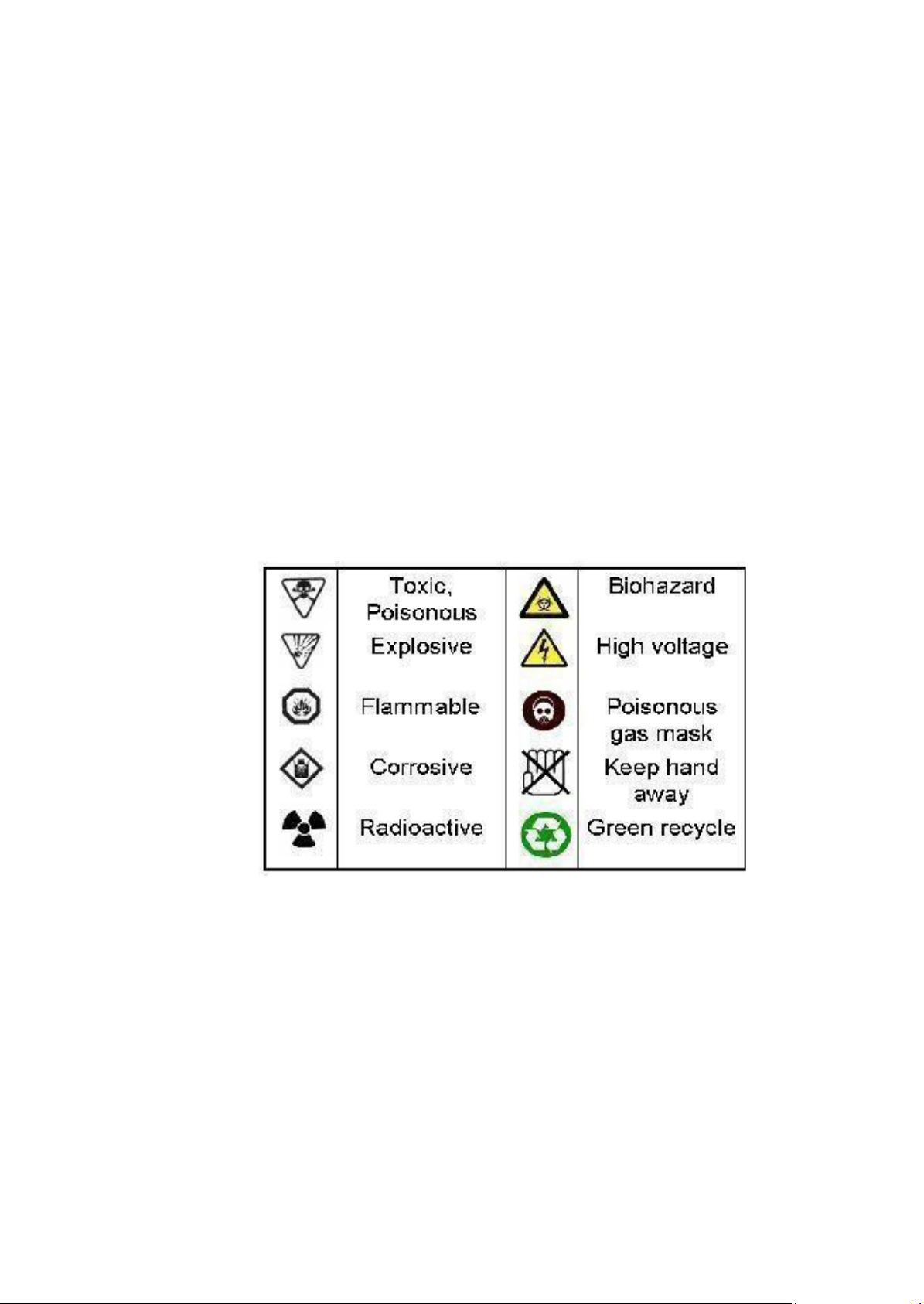
Know the warning symbols for specific chemical hazards (see below). 8.
Use fume cupboard for hazardous chemicals. 9.
Work in a logical, tidy manner and minimize risks by planning in advance.
10. Clean up working bench and experimental tools at the end of each lab session.
11. Dispose wastes in appropriate containers.
Practical 1: MICROSCOPY AND CELL OBSERVATION
AIMS OF THE PRACTICAL:
Familiarize students with the use and care of microscope, an indispensible instrument for
anyone who works in biological fields.
Introduce students the microscopic sample preparation of plant cells and animal cells. lOMoAR cPSD| 59078336 1. MICROSCOPY
Microscopy is defined as a study of using microscopes to
observe very small objects that are invisible to the human naked eye.
Light microscope can enhance our capacity to view detail by 1000
times, so that we can see samples as small as 0.1 μm in diameter.
High-tech microscopes, such as Transmission Electron Microscope
(TEM) and Scanning Electron Microscope (SEM), are able to give
visual magnification up to 200,000 times and fascinating features in
comparison with unaided visibility. In fact, the biological
understanding of cell structures and functions would be extremely
restricted without microscopes.
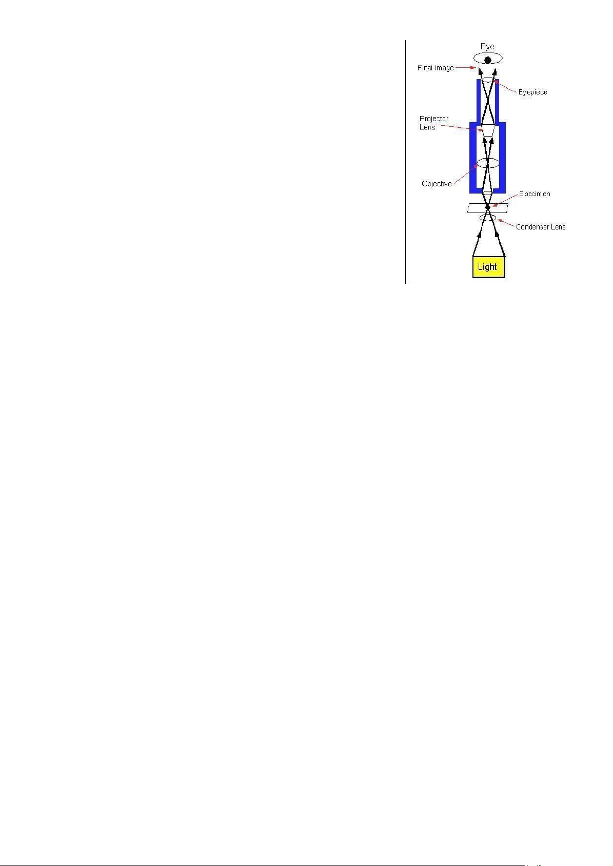
All microscopes consist of a coordinated system of lenses arranged so that a magnified image of the specimen can be seen by
Figure 1.1: Basic principle of
the viewer. The main differences among different types of microscopes
are the power source to produce the picture a light microscope or optical microscope.
and the arrangement of the lens system and maximal resolution (resovling power) they can offer. In
general, microscope works base on the magnification of specimen image through a series of lenses.
There are different types of microscope: Compound light microscopes, Stereoscopes, Confocal
microscopes, Scanning electron microscopes, Transmission electron microscopes…
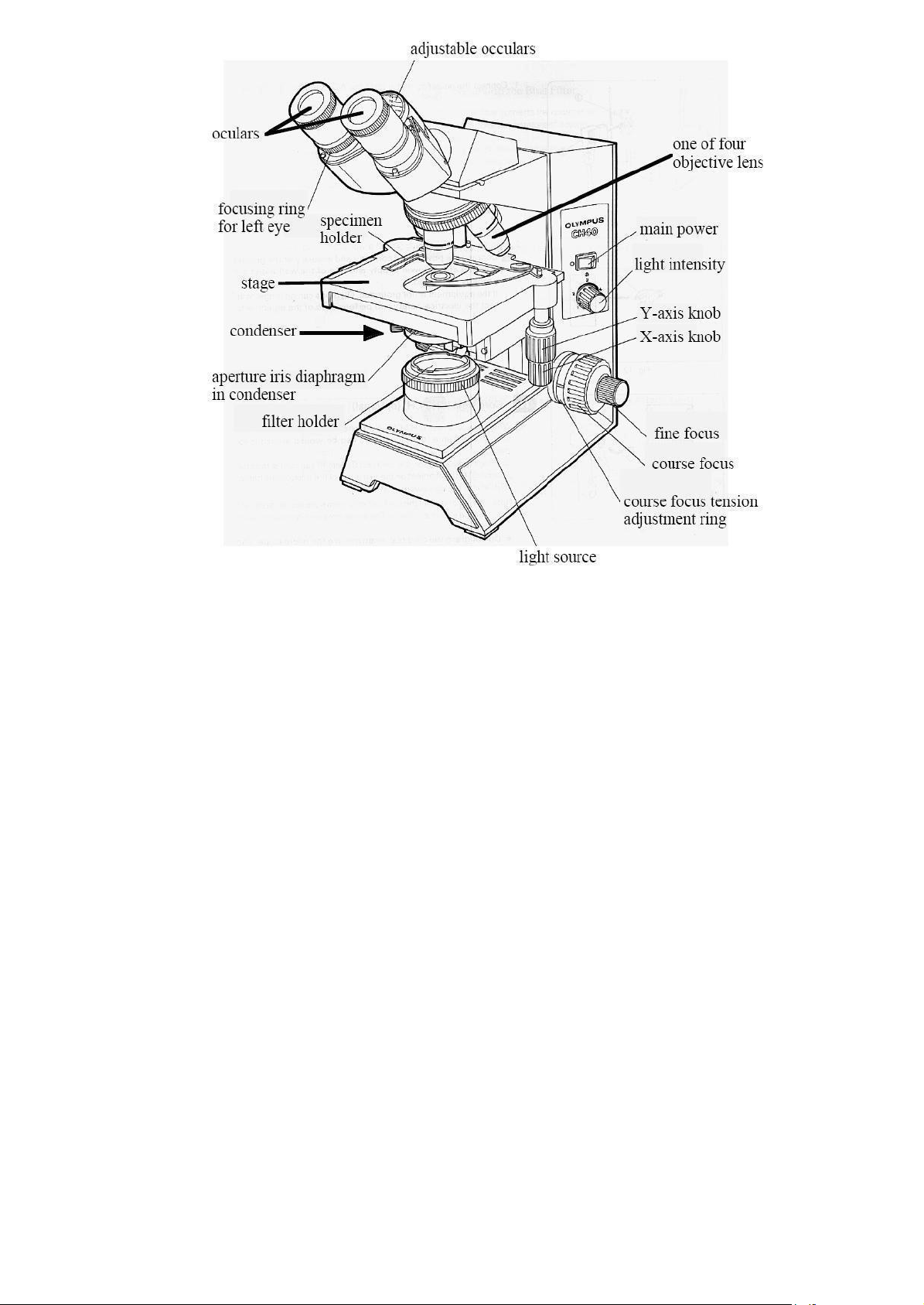
Construction of a compound Microscope
Eyepieces (ocular lens): are placed at the top of the body tube. They transmit and magnify the
image from the objective lens to eyes. Eyepiece has its own magnification power. The eyepieces that you
use will have magnification power of 10 (10x).
Objective lens: are lens or series of lenses that gather light from specimen and help to magnify
the specimen image. This is the most important part of a microscope. To figure the total magnification
of an image that you are viewing through the microscope, take the power of the objective lens (4x, 10x,
40x, 100x) and multiply by the power of the eyepiece. lOMoAR cPSD| 59078336
Figure 1.2: Different parts of a compound light microscope.
Body tube: allows light to travel from the objectives through a series of magnifying lenses to
the eyepieces. In your microscope, the eyepieces are held at an angle for convenient use, and the body
tube contains a prism that bends the light rays so that they will pass through the eyepieces.
Stage: is the surface or platform on which you can place your specimen. Stage clips or
specimen holder is used to clamp your specimen on the stage. For your microscope, the stage is movable
and is called a mechanical stage. The movement is controlled by two knobs (X-axis and Y- axis knobs)
located on the bottom of the stage. They allow the specimen to move vertically and horizontally.
Condenser: is a lens system under the stage that gathers light from the light source and focuses it onto the specimen.
Condenser adjustment control: is to adjust the height of the condenser. Usually, the
condenser always will be all the way up.
Aperture iris diaphragm: is to control the level of light that can go through the condenser.
Light source: the illuminator of most microscopes is built into the base of the microscope and
controlled by on/off switch. You can control the light intensity by adjusting the voltage of a transformer attached to the illuminator.
Coarse focus knob and fine focus knob: You focus a microscope by using the Coarse and
Fine Focus knobs. Both coarse (large) and fine (small) adjustment knobs are found on both sidesof our
microscopes. Remember that the coarse adjustment is used only with the low-power objective (4x). These
knobs control a gear mechanism that raise and lower the stage. lOMoAR cPSD| 59078336
Proper Use of a Compound Microscope
1.1. General instructions
Avoid dropping the microscope, banging it against a lab bench, or having the eyepieces fall out.
- Carry the microscope upright using both hands. Keep one hand on the arm and another at the bottom of the microscope.
- Keep microscope away from the edge of the bench.
- Switch off the illuminator and remove power cords from the power supply when not in use.
Avoid breaking a coverslip and microscope slide, and even the objective lens while focusing.
- First adjust the stage to the lowest position.
- Locate the ready specimen using the lowest power objective lens, and then switch to the higher power objective lenses.
- Never focus the high power lenses with the coarse adjustment knob, and never use these lenses to examine thick specimens. 1.2. Focusing
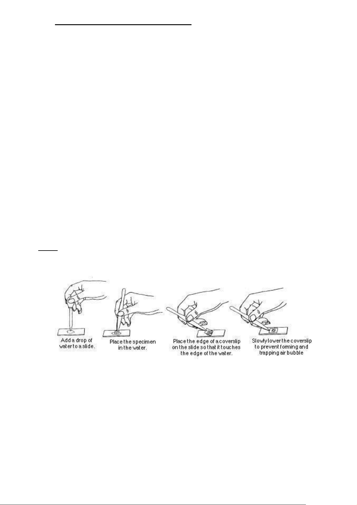
Notice: Always use clean microscope slides, and proceed from the lowest power to the highest power objectives.
Figure 1.3. Preparing a wet mount slide
1. Clean the eyepieces and objective lens using lens-cleansing papers (if necessary)
2. Cut out a letter “e” from a newspaper or other printed page. Clean a microscope slide and
prepare a wet mount of the letter, following Figure 1.3.
3. Put the lowest power objective (4x) in position, lowest the stage of the microscope, and place
the slide on the microscopic stage.
4. Switch on the illuminator and diaphragm fully, and adjust the condenser level appropriately.
5. Move the specimen into the bright area on the stage using X and Y-axis knobs. lOMoAR cPSD| 59078336
6. Lift the stage up close to the objective lens using the coarse focus (clock-wise direction) while
observing through the eyepieces until the specimen comes into focus.
7. Use the diaphragm to adjust the light intensity as necessary as well as center the specimen by
moving the slide using X and Y-axis knobs.
8. Switch from the scanning objective (4x) into the high-power objective (10x). Refine the focus
by gentle adjustment with fine focus knob only.
9. Switch to the higher-power objective (40x or 100x) and again adjust the focus with the fine focus knob only. 2. CELL OBSERVATION
Objective: Observation different cell types (plant cells and animal cells) under microscope at different objective lens.
Materials and Equipments: • Onion bulb • Lugol solution • Light microscope • Blade • Glass slides • Forceps • Coverslips • Tooth-pick
• Distilled water • Scissors
• Pasteur pipette • Tissue paper lOMoAR cPSD| 59078336
2.1. Plant Cells – Onion Epidermis Cells 1.
Remove a piece of an onion leaf from a section of an onion bulb. 2.
Break the piece of onion leaf into half as shown in Figure 1.4. The outer epidermis layer
should be easy to separate from the rest of the leaf.
Figure 1.4. How to obtain a piece of onion for slide preparation 3.
Place the epidermis layer flat on a slide. Wrinkles will trap air bubbles and obscure your observations. 4.
Add a drop of distilled water/Lugol solution and cover with a coverslip. 5.
Excess distilled water/Lugol solution should be removed using tissue paper. 6.
Observe your slide with your microscope and take photo. Remember to locate a good
region of the epidermis with the lowest magnification (4x) lens before observing details of cell
structure with higher magnifications up to 10x and 40x.
2.2. Animal Cells – Human Cheek Cell Epithelium
1. Gently scrape the inside of your cheek with the broad end of a toothpick. (You won’t need to
puncture your cheek to obtain a good supply of cells.)
2. Smear your cheek scrapings on a clean slide. Wrap your toothpick in a dry paper towel and
immediately dispose it into the waste-container provided.
3. Make a wet mount of your cheek cells by adding a drop of Lugol solution to the slide. Excess
Lugol solution should be removed using tissue paper.
4. Add a coverslip and observe the slide at very low light intensity. It helps you to observe the cells easily.
5. When you locate some cheek cells, at 4x objective lense center them in the field of view and
move to the next power level (10x) for observation. Re-focus and center your cheek cells and then view
them with the high power (40x) objective lens. Observation and take photo. lOMoAR cPSD| 59078336
Practical 2: ORGANIC COMPOSITIONS OF THE CELL PRE-LABS 1.
What are 4 classes of biological macromolecules and their building blocks? 2.
Describe structure of carbohydrate (starch, sugar). 3.
What is the difference between Lugol and Iodine solution? How can we prepare them? 4.
Describe structure of protein. 5.
How would you prepare 100 ml of 0.5% CuSO4 solution from CuSO4.5H2O (MW = 250)? 6.
Where can we find lipid in plant cells and animal cells? 7.
Describe structure of nucleic acid. 8.
In the forthcoming practical session, you will have to use a number of different
chemical solutions: Lugol solution, concentrated HCl, NaOH, CuSO4, soudan III, 20% ethanol and
glycerin. List three solutions, which are most potentially toxic and thus require caution while handling, in your opinion. Explain your reason.
AIMS OF THE PRACTICAL:
Help students to identify the three main organic compositions of the cell: carbohydrates, proteins
and lipids on different samples. 1. CARBOHYDRATES
Carbohydrates make up a group of chemical compound found in plant and animal cells. They have
the empirical formula CnH2nOn or (CH2O)n. An empirical formula of carbohydrate tells the atomic
compositions of the compound. Carbohydrates are divided into 4 groups: monosaccharides,
disaccharides, oligosaccharides and polysaccharides. Most monosaccharides, or simple sugars, are
found in grapes, other fruits, and honey. Disaccharides include sucrose, lactose and maltose; the
disaccharide sucrose can be found in sugar beets and cane sugar. Disaccharides are composed of two
monosaccharide units linked together by a glycosidic bond. Oligosaccharides, which consist of three to
six monosaccharide units, are rather infrequently found in natural sources (bamboo shoots, vegetables,
onion, wheat, etc.). Polysaccharides (the term means many sugars) represent most of the structural and
energy-reserve carbohydrates found in nature. Large molecules may consist of as many as 10,000
monosaccharide units linked together, polysaccharides vary considerably in size, in structural complexity,
and in sugar content (such as cellulose, starch, glycogen). Oligosaccharides and polysaccharides are also
linked together by glycosidic bonds. lOMoAR cPSD| 59078336
Glucose (carbohydrate) is the primary products of plant photosynthesis. The simplified light-
driven reaction of photosynthesis results in the formation of carbohydrates:
nH2O + nCO2 (CH2O)n + nCO2.
This type of carbohydrate is found in the structure of plants and is used in the reverse reactions of
photosynthesis (respiration) or is consumed as fuels by plants and animals. The excess glucose is stored
in form of starch in plants. While in animal, the excess produced glucose is stored in form of glycogen in liver and muscles.
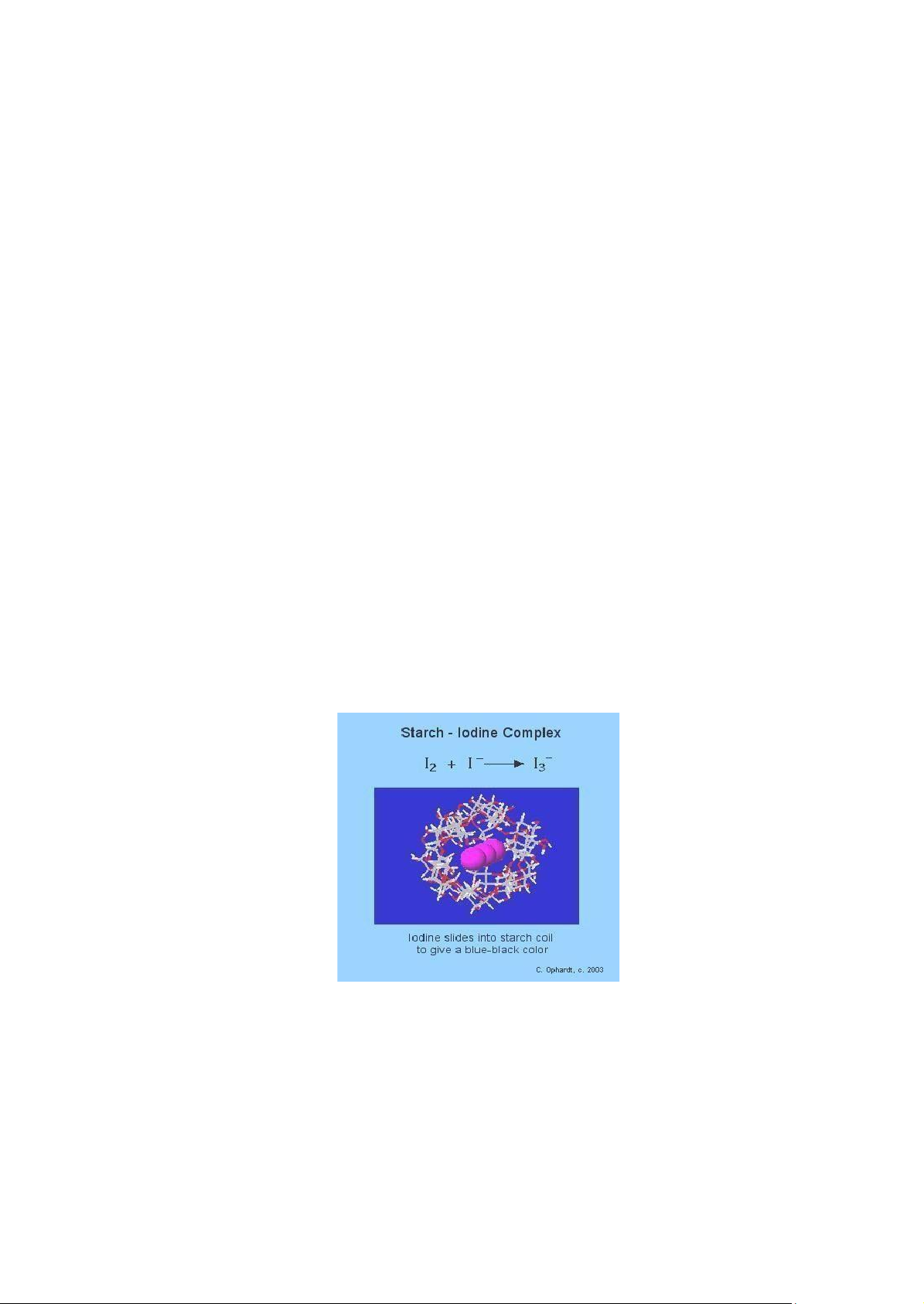
The most stable three-dimensional structure for starch and glycogen is a tightly coiled helix
stabilized by inter-chain hydrogen bonds.
In amylose (with no branches) this structure is regular enough to allow crystallization and thus
determination of the structure by X-ray diffraction. Each residue along the amylose chain forms a 60o angle
with the preceding residue, so the helical structure has six residues per turn. For amylose, the core of the
helix is of precisely the right dimensions to accommodate iodine in the form I3- or I5- (iodide ions), this
complex has the deep blue color and this interaction with iodine is a common qualitative test for amylose.
However, this glycosidic bond is easily wrecked by heat, enzyme, acidic or basic treatment.
Hydrolyzing the starch with acid or exposing to high temperature result in the formation of mixture of
saccharides with different lengths (including the one-, two- or number of glucose chains). Once the helix
structure of homopolycarbohydrate is broken, the conformation with iodine no longer exists.
Figure 2.1: Interaction between amylose and iodine ions when starch is treated with Lugol solution
Objective: How to detect the starch in the sample/solution by Lugol solution. And understand the
effect of temperature to the structure of the starch.
Materials and equipments:
• Potato, rice starch suspension, microscope, glass slides, coverslips
• 1 test tube, test tube racks, test tube clamps, Lugol solution, knife, tooth-pick
• Pasteur pipette, 2M HCl solution, waterbath, distilled water, tissue paper lOMoAR cPSD| 59078336
1.1. Task 1 – Microscopic Observation of Starch Granules
1. Prepare the clean glass slide and coverslip.
2. Cut the potato and scratch potato at the edge of this cut.
3. Collect the scratching and place on the slide, add a drop of distilled water and cover with
coverslip. Excess distilled water should be removed using tissue paper.
4. Observe under microscope 4x and 10x. Please take photo.
5. After that, adding 1 drop of Lugol solution to the edge of the coverslip, observe the color
change and please do not use microscope for observation. Please take photo, too.
1.2. Task 2 – Chemical detection of starch
1. Add 5ml of starch suspension into a test tube.
2. Then, take out a drop of rice starch suspension and put onto the glass slide. Add 1 drop of Lugol
solution and mix well using a toothpick. Then, please put another drop of Lugol soultion on the same
glass side. Observe the color change of the mixture (compare with the original color of Lugol solution). Please take photo.
3. After that, add 5 drops of 2M HCl solution into that test tube containing 5ml of starch suspension. Mix well.
4. Take out 1 drop of starch-HCl mixture onto another glass slide and test the color with 1 drop
of Lugol solution. Please take photo.
5. Next step is to place the test tube containing starch and HCl into the rack which has been
already submerged in the hot water (the waterbath is set to 100oC) and boil this suspension. Every 2
minutes, take out one drop of hydrolysed starch-HCl mixture using pateur pipette and put onto another
clean glass slide and add 1 drop of Lugol solution, mix well using tooth-pick. Observe the change of color intensity
and take photo.
6. Continue to boil and test with Lugol solution until no color change is detected. Mark the time
that the color does not change. 2. PROTEINS
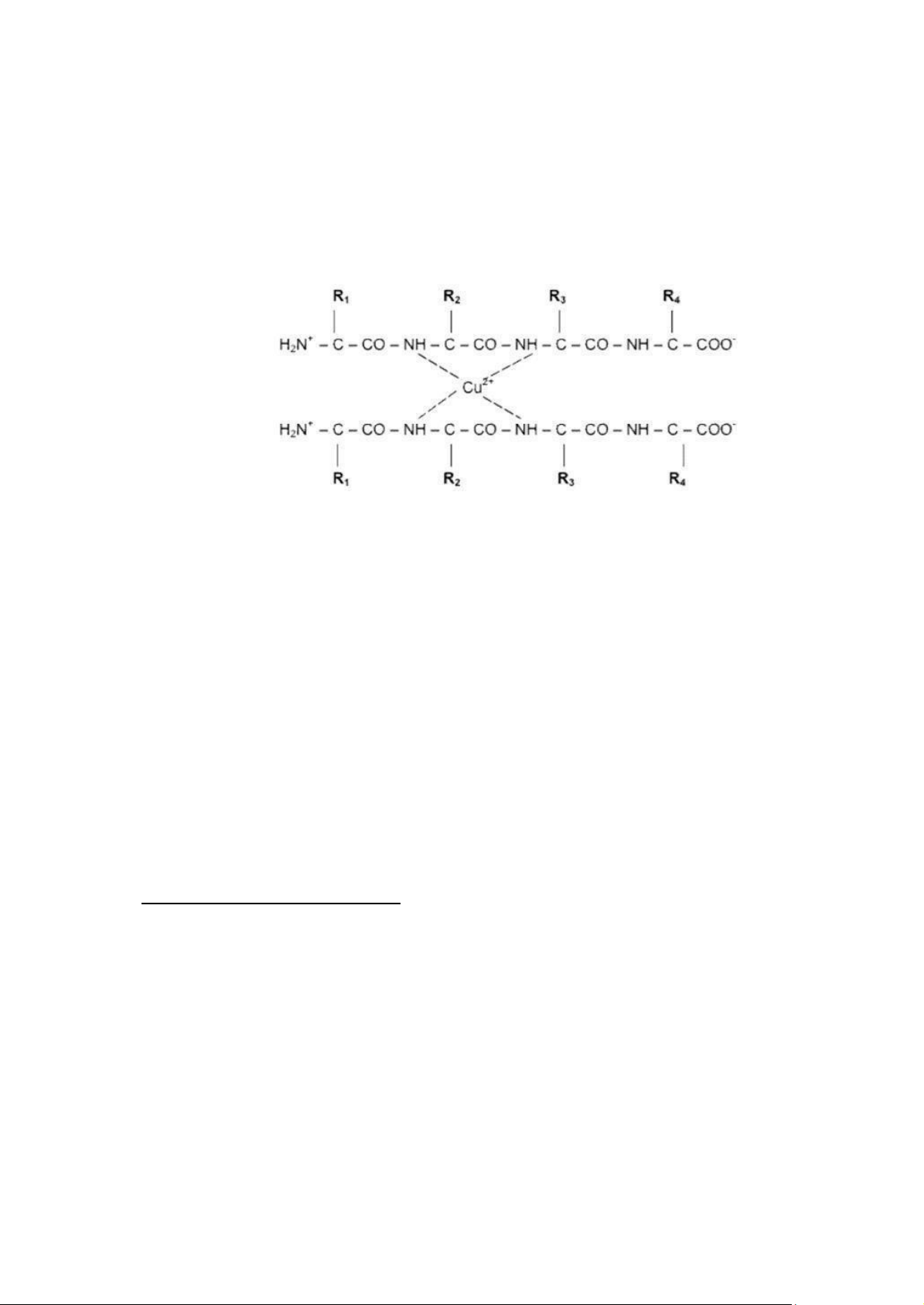
Proteins are complex polymers composed of amino
acids. Amino acids contain carbon, hydrogen, oxygen,
nitrogen and sometimes sulfur and serve as monomers for
making peptides and proteins. Amino acids are linked
together by peptide bonds in which the carboxyl carbon of
Figure 2.2: A peptide bond links two
one amino acid forms a covalent bond with the amino amino acid residues nitrogen of the other amino acid. lOMoAR cPSD| 59078336
Short chains of amino acid are called peptides, longer chains of amino acids are called
oligopeptides or polypeptides.
In the strong basic environment, two nitrogen atoms from two adjacent peptide bonds can
coordinate with other two nitrogen atoms on other peptide chain in the conformation with metal ions
such as Cu, Zn… if present. The complex gives out the color ranging from purple to red depending on
the length of peptide chain (or the number of peptide bonds). The Biuret reaction is used to detect the
presence of peptide or protein in sample.
Figure 2.3. Protein-Copper complex formation in Biuret reaction
Objective: Understand the Biuret reaction and the role of chemical reagents in protein detection.
Materials and Equipment:
• Protein suspensions (egg white and cow milk)
• 2 test tubes, test tube rack • Pasteur pipette • 10% NaOH solution • 0.5% CuSO4 solution
Qualitative Detection of Proteins
1. We need 2 test tubes, each test tube containing 2 ml of protein suspension.
2. Add 2 ml (or 10 drops) of 10% NaOH solution. Mix well.
3. Add 3 drops of 0.5% CuSO4 solution into the test tube.
4. Observe the color and take photo of 2 test tubes. 3. LIPIDS
Lipids are a group of naturally occurring molecules that include fats, waxes, sterols, fat-soluble
vitamins (such as vitamins A, D, E, and K), monoglycerides, diglycerides, triglycerides, phospholipids,
and others. Biological lipids originate entirely or in part from two distinct types of biochemical subunits
or "building-blocks": ketoacyl and isoprene groups. Using this approach, lipids may bedivided into eight lOMoAR cPSD| 59078336
categories: fatty acids, glycerolipids, glycerophospholipids, sphingolipids, saccharolipids, and
polyketides (derived from condensation of ketoacyl subunits); and sterol lipids and prenol lipids (derived
from condensation of isoprene subunits).
The main biological functions of lipids include storing energy, signaling, and acting as structural
components of cell membranes. Lipids may be broadly defined as hydrophobic or amphiphilic small
molecules; the amphiphilic nature of some lipids allows them to form structures such as vesicles,
liposomes, or membranes in an aqueous environment.
Objective: Detect and observe the lipid in plant cells using red Soudan III solution.
Materials and Equipments: • • Peanut (soaked in water) Microscope
• Soudan III solution • Glass slide and coverslip
• 20% Ethanol • Pasteur pipette • Blades, distilled water • Tissue paper, knife
Qualitative detection of lipids
1. Scratch the peanut by knife (which was soaked in water).
2. Place it on glass slide; add a drop of Soudan III solution. Keep sample in this solution for staining in 10 minutes.
3. Wash the slide with 20% ethanol.
4. Add a drop of distilled water to the sample then put the coverslip on. Excess distilled water
should be removed using tissue paper.
5. Observe the lipid granules stained in peanut cells using microscope 40x. Please take photo.
Practical 3: PHOTOSYNTHESIS AND TRANSPIRATION PRE-LABS
1. What are autotrophs and heterotrophs? Give each one example.
2. What is “photosynthesis”? How many stages are there in the photosynthesis? Describe.
3. Where is the chlorophyll distributed in plants and animals?
4. What is the function of chlorophyll?
5. Define the terms “transpiration” and “respiration”. What is the difference between them?
6. What are stomata and guardcells? Describe their distribution on the leaf. lOMoAR cPSD| 59078336
AIMS OF THE PRACTICAL:
Help students to understand the process of photosynthesis by examining the products of photosynthetic reaction. 1. PHOTOSYNTHESIS
Photosynthesis is the process by which plants, some bacteria, and some protistans use the energy
from sunlight to produce sugar, which can then be converted into ATP, the "fuel" used by all living things
via cellular respiration process. The overall photosynthesis reaction can be summarized asfollow:
6H2O + 6CO2 C6H12O6 + 6O2
Photosynthesis is associated with the activities of
the green pigment chlorophyll. Several modifications of
chlorophyll occur among plants and other autotrophic
organisms. A pigment is defined as a substance that can
absorb light. The color of the pigment comes from the
wavelengths of light reflected (in other
words, those are not absorbed). Chlorophyll, the green pigment common to all photosynthetic cells, absorbs all wavelengths of
Figure 3.1: Structure of a
chloroplast visible light except green, which reflects the green colour to be seen by our eyes.
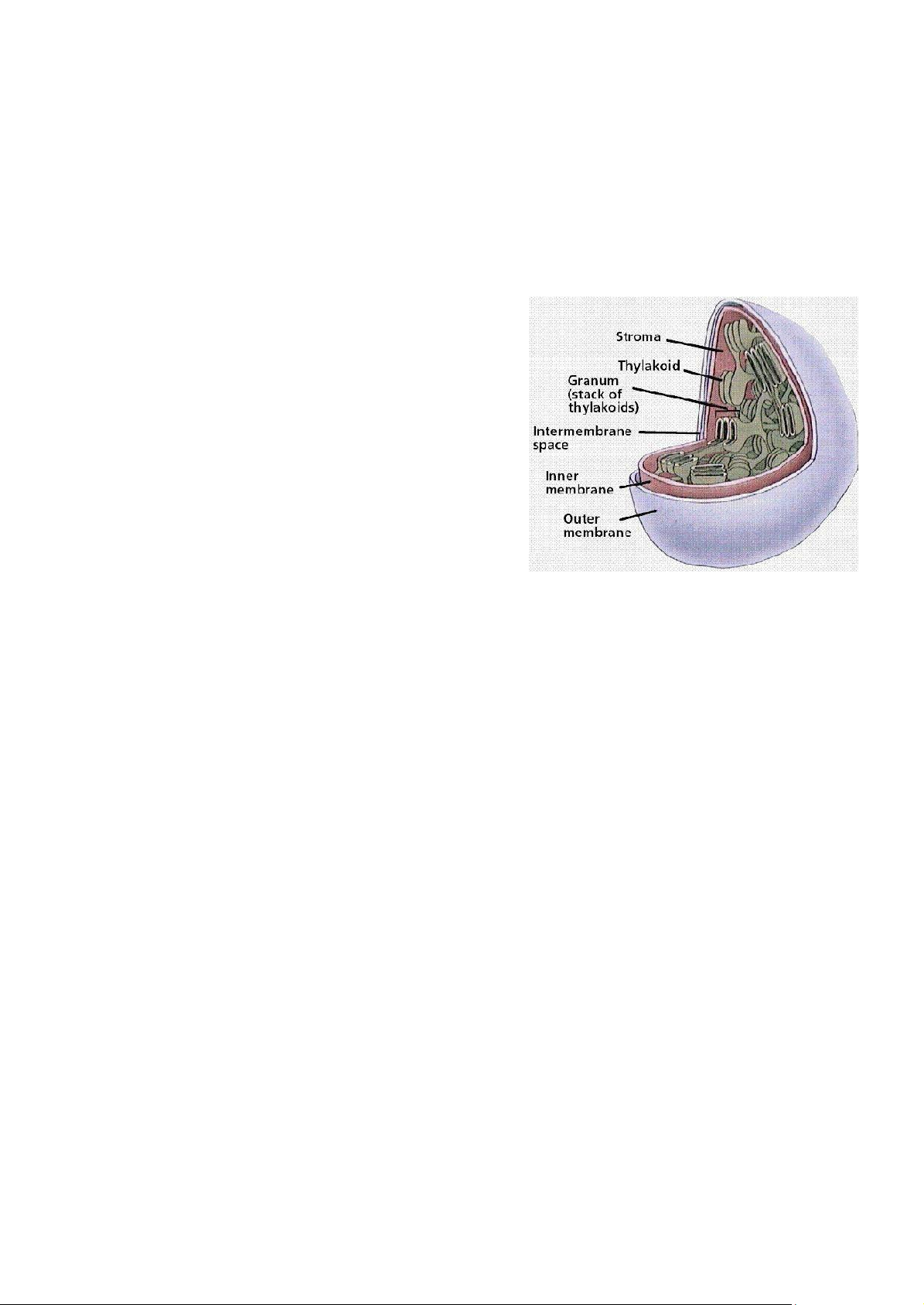
Chlorophyll is embedded in the membrane of a specific structure called thylakoid. This is the unit
of photosynthesis. Thylakoids are stacked like pancakes in stacks known collectively as granum.
The areas between grana are referred as stroma. While the mitochondrion has double membrane systems,
the chloroplast has three, forming three compartments.
Objective: In this experiment, we will examine the presence of photosynthetic products in plant.
Materials and Equipments • Waterweed branches
• Leaves with covered part (you have already prepared at home) • Waterbath
• Ethanol 70%, Lugol solution
• Beaker, funnel, 3 test tubes, test tube rack, test tube clamps • Petri dish, forceps, tissue paper, foil paper lOMoAR cPSD| 59078336
Task 1 – Examination of Oxygen Formation 1.
Prepare one beaker with two thirds of water. 2.
Turn down the funnel to the beaker. 3.
Put 3 waterweed branches into the funnel. 4.
Cover the tunnel end with the test tube filled with water and bring this beaker to the
light condition (near the window). 5.
Observe the formation of coming up bubbles and record the level of water goes down
every 5 days up to 15 days. Take photo please. 6.
After the incubation time, take out the test tube from the funnel while still keep it at the
original orientation, cover the test tube by hand and upturn this test tube. 7.
Remove hand and immediately test the gas in test tube with the burned match. 8.
Observe what happens with the fire from match.
1.1. Task 2 – Examination of Starch Formation 1.
Choose one leaf on a growing tree. Clean this leaf with water and tissue paper and then dry. 2.
Use a piece of black paper or cotton to cover the middle half of this leaf, make sure that
no light can penetrate to this part. Leave it for at least two weeks. (*should not use electric tape –
too stick to remove without breaking the leaf) 3.
On the day of doing the experiment, pick this leaf from the tree. 4.
Remove the cover (paper or cotton). Notice the color difference between two areas of
the leaf and take photo. 5.
Put this leaf into the boiling water (waterbath) for 5 minutes. 6.
Use forceps to take out the leaf from hot water and put it into the test tube with ethanol
70%, put this test tube with leaf into boiling water in the rack, continue to boil until the green color disappears. 7.
Take out the leaf from test tube, wash with water and dry the boiled leaf with tissue
paper. Then, stretch it out on a petri dish and then add Lugol solution into the dish. 8.
Observe the color in 2 areas of the leaf. Take photo please. 2. TRANSPIRATION
Water enters the root and is transported up to the leaves through specialized plant cells known as
xylem. Land plants must guard against drying out (desiccation), mainly via the leave system. Adaptation
through evolution has made the leaf surface usually covered by a cuticle layer in order to avoid water
evaporation. However, the leaves also contain specialized structures known as stomata to allow gas to lOMoAR cPSD| 59078336
enter and leave the leaf. This is especially important for photosynthesis process. Carbon dioxide cannot
pass through the protective waxy layer covering the leaf (cuticle), but it can enter the leaf through an
opening the stoma flanked by two guard cells. Likewise, oxygen produced during photosynthesis can
only pass out of the leaf through the opened stomata. Unfortunately for the plant, while these gases are
moving between the inside and outside of the leaf, a great deal water is also lost though the same way.
Cottonwood trees, for example, will lose 100 gallons of water per hour during hot, sunny days.
Objective: We will examine the evaporation of water in leaf by using the indicator from Co2+. The
Co2+ ion can form the complex with absorbed water and change its original color (blue) to the color of
its conjugate with water (pink). CoCl 2- 2+ 4 + 6H O 2 Co(H O) 2 + 4Cl- blue pink
Materials and Equipments
• One healthy leaf, 3% CoCl2 solution
• Absorbent paper (2 pieces/group) • Scissors, Desiccator • Tissue paper, petri dish • Sticking tape, forceps
Examination of water loss on leaf surface
1. Cut the absorbent paper into pieces with size smaller than size of sticking tape and of leaves examined.
2. Soak these paper into 3% CoCl2 solution for two minutes and then dry the paper at 80oC in
desiccator till dried. Each group will need 2 pieces of CoCl2 paper. Notice the color of paper and take photo.
3. Clean and dry one healthy leaf with tissue paper (make sure that the leaf is completely dry). lOMoAR cPSD| 59078336
4. Use forceps to take out the dried pieces of CoCl2 paper and place them on a sticking tape.
5. Quickly and tightly apply these tapes onto two surfaces of leaf at the same time (upper and lower
surfaces). You need to ensure that no moisture from the air can enter to the CoCl2 paper. Then,
take photo as observation time of 0 min.
6. Check the color change of paper on both surfaces of leaf after every 5 mins and notice the time
needed for this color change. Repeat this step until 30 mins. During the experiment, you need to
take photo as indicated time.
Practical 4: OSMOSIS AND LEAF STOMATA PRE-LABS
1. What does sodium chloride (NaCl) do in osmosis process?
2. After adding hypertonic solution (5% NaCl), plant/animal cells decrease their size during
osmosis process. Could you explain this phenomenon?
3. What will happen when plant/animal cells are in contact with isotonic solution (0.85% NaCl)? lOMoAR cPSD| 59078336
4. Are the stomata necessary for plants? Why?
5. What will the leaf stomata respond when plants lack of water?
AIMS OF THE PRACTICAL:
Examine the osmosis process in plant cells.
Observe the leaf stomata on the both surfaces of plant cells. 1. OSMOSIS
Osmosis is the process where water molecules flow across a differentially permeable membrane
from a solution with a lower solute concentration to a solution with a higher solute concentration. This
membrane allows only certain types of molecules to pass through it or permeate it freely. Cellular
membrane is a type of differentially permeable membrane.
Osmosis does not require expenditure of energy but an energetically "downhill" process. Since the
water must lose energy as it moves by osmosis, water must move from an area of greater water potential
to an area of lower water potential:
• Hypertonic solution has water potential outside the cell lower than inside the cell, then there
will be a net movement of water out of the cell.
• Hypotonic solution has water potential outside the cell greater than inside the cell, then osmosis
will be a spontaneous net movement of water into the cell.
• In isotonic solution, the water potential on each side of a cell membrane is the same, there will
be no net movement of water across the membrane.
Osmotic pressure of a solution is proportional to the effective concentration of dissolved particles,
regardless of the size or chemical nature of the particle.
Objective: Demontrate and observe the osmosis and osmotic pressure using epidemic plant cells from
Tradescantia spathacea leaf.
Materials and Equipments
• Tradescantia spathacea leaf
• Distilled water (0% NaCl - sodium chloride solution) hypotonic solution
• 5% NaCl (sodium chloride) solution hypertonic solution
• 0.85% NaCl (sodium chloride) solution isotonic solution
• Glass slides, coverslips, forceps, microscope, tissue paper, scissors
Plant cells – Plasmolysis
Plant cells always have a strong cell wall surrounding them. When taking up water by osmosis they
start to swell, but the cell wall prevents them from bursting. Plant cells become "turgid" when they are
put in dilute solutions. Turgid means swollen and hard. The pressure inside the cell rises; eventually the lOMoAR cPSD| 59078336
internal pressure of the cell is so high that no more water can enter the cell. This liquidor hydrostatic
pressure works against osmosis. Turgidity is very important to plants because this iswhat make the green
parts of the plant "stand up" into the sunlight.
When plant cells are placed in concentrated NaCl solutions they lose water by osmosis and they
become "flaccid"; the exact opposite of "turgid". If you put plant cells into concentrated NaCl solutions
and look at them under a microscope you would see that the contents of the cells have shrunkand pulled
away from the cell wall: they are said to be plasmolysed. And this phenomenon is call plasmolysis.
When plant cells are placed in a solution which has exactly the same osmotic strength as the cells
they are in a state between turgidity and flaccidity. We call this incipient plasmolysis. "incipient" means
"about to be". When one forgets to water the potted plants, their leaves drop. Although their cells are not
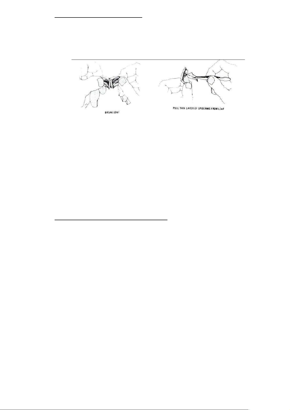
plasmolysed, they are not turgid and so they do not hold the leaves up into the sunlight. Procedure 1.
Break Tradescantia spathacea leaf into half to easily separate from the rest of the leaf.
You need to collect a thin epidermis layer (purple side) of the leaf. Prepare 3 glass slides for 3 specimens. 2.
In each specimen, add a drop of 0.85% NaCl, 5% NaCl or distilled water on a clean glass slide. 3.
Place the purple layer to the saline on the slide. Add a coverslip. 4.
Excess saline solution should be removed using tissue paper. 5.
Examine the plant cells with the lowest power lens (4x) first, after observing the plant
cells, you use the next power level (10x and 40x) for observation. 6.
Observe the plant cells and the difference among samples (focus to the cellular content
- purple area) that occur as the solutions reach them. Please take photo at objective lens 40x.
3.1. Animal cells – Hemolysis
The cell membrane of erythrocytes (red blood cells) is permeable to water but relativelyimpermeable to
salts. If red blood cells are placed in an isotonic saline solution (0.85% NaCl), the cells will retain their shape and size.
If the red blood cells are in a hypotonic saline solution, water will enters the cells more rapidly than it
leaves. As a consequence, the red blood cells swell and ultimately bust, releasing hemoglobin. This
phenomenon is called hemolysis. Red blood cells if placed in a hypertonic saline solution will shrink and
appear to have a bumpy, irregular outline. The cells are said to be crenated. 2. LEAF STOMATA
Stomata, any of the microscopic openings or pores in the epidermis of leaves. They can also occur on
young stems but less commonly on leaves. Stomata are generally more numerous on the underside of leaves. lOMoAR cPSD| 59078336
They provide for the exchange of gases between the outside air and the branched system of interconnecting air canals within the leaf.
A stoma opens and closes in response to the internal pressure of two sausage-shaped guard cells that
surround it. The inner wall of a guard cell is thicker than the outer wall. When the guard cell is filled with
water and it becomes turgid, the outer wall balloons outward, drawing the inner wall with it and causing the stoma to enlarge.
Guard cells work to control excessive water loss, closing on hot, dry, or windy days and opening when
conditions are more favourable for gas exchange. For most plants, dawn triggers a sudden increase in stomatal
opening, reaching a maximum near noon, which is followed by a decline because of water loss. Recovery
and reopening are then followed by another decline as darkness approaches. In plants that photosynthesize
with the CAM carbon fixation pathway, such as bromeliads and members of the family Crassulaceae, stomata
are opened at night to reduce water loss from evapotranspiration.
The main functions of stomata:
1. Gaseous exchange- Stomatal opening and closure help in the gaseous exchange between the plant and surrounding.
2. It helps in transpiration and removal of excess water in the form of water vapour.
3. Stomatal closure at night prevents water from escaping through pores.
4. It maintains the moisture balance according to weather by opening and closing.
5. Stomata facilitate carbon dioxide uptake and release of oxygen during the process of photosynthesis.
Objective: To observe leaf stomata on the upper and lower surfaces of plant cells from Tradescantia
spathacea monocot leaf and paper flower (Bougainvillea spectabilis) dicot leaf.
Materials and Equipments:
• Tradescantia spathacea monocot leaf and paper flower (Bougainvillea spectabilis) dicot leaf • Distilled water
• Glass slides, coverslips, forceps, clear nail polish
• Microscope, tissue paper, scissors Procedure
1. Pick a healthy leaf from the potted plant.
2. Paint a swath of the nail polish on either the upper and lower surfaces of the leaf.
3. Try to avoid the veins if possible as they can make the polish harder to deal with later.
4. Let the nail polish patch dry for a couple of minutes.
Tài liệu liên quan:
-
Final Exam Notes - Genetic Variability & Development | Môn Biology - Trường Đại học Quốc tế, Đại học Quốc gia Thành phố Hồ Chí Minh
127 64 -
Lab Report 4: Enzyme Activity of Amylase and Catalase in Biology | Môn Biology - Trường Đại học Quốc tế, Đại học Quốc gia Thành phố Hồ Chí Minh
105 53 -

Lecture 5: Understanding Genetics I - Cell Cycle & Mitosis | Môn Biology - Trường Đại học Quốc tế, Đại học Quốc gia Thành phố Hồ Chí Minh
117 59 -
Final review | Môn Biology - Trường Đại học Quốc tế, Đại học Quốc gia Thành phố Hồ Chí Minh
127 64