Final review | Môn Biology - Trường Đại học Quốc tế, Đại học Quốc gia Thành phố Hồ Chí Minh
Bacteria, the oldest known life forms on Earth, belong to the prokaryotic group. These single-celled organisms, which have existed for billions of years, lack a nucleus and have evolved into a diverse range of types that can adapt to various environments. Tài liệu được sưu tầm gồm 7 trang, giúp bạn ôn tập tốt hơn. Mời các bạn đón xem.
Môn: Biology 10 tài liệu
Trường: Trường Đại học Quốc tế, Đại học Quốc gia Thành phố Hồ Chí Minh 2 K tài liệu
Tác giả:


Preview text:
lOMoAR cPSD| 59078336
- Bạn nghĩ gì về Vi khuẩn(Tự viết Review)- Slide thì là Chap 10
Bacteria, the oldest known life forms on Earth, belong to the prokaryotic group.
These single-celled organisms, which have existed for billions of years, lack a
nucleus and have evolved into a diverse range of types that can adapt to various
environments. They can thrive in the human body, arctic snow, soil, rocks, and even the deep ocean.
Some bacteria live symbiotically with plants and animals, including humans,
while others reside in soil or on decaying plant matter, playing a crucial role in
nutrient cycling. Although some bacteria cause food spoilage and crop damage,
many are beneficial, such as those used in producing fermented foods like yogurt
and soy sauce. Only a small percentage of bacteria are pathogenic, causing
diseases in plants and animals. Size+Structure
Bacteria are microscopic, with sizes ranging from 1 to 10 micrometers in length
and typically 1 to 2 micrometers in diameter. Their structure includes several key
components. The cell wall, made of peptidoglycan, maintains the cell's shape,
provides protection, and prevents bursting in hypotonic environments. The plasma
membrane, composed of phospholipids, proteins, and carbohydrates, functions in
transport, biosynthesis, and energy transduction. The cytoplasm aids in cellular
growth, metabolism, and replication, housing essential chemicals and nutrients.
Ribosomes are the sites of protein synthesis, while plasmids, small circles of DNA,
facilitate genetic exchange between cells. Flagella and pili aid in movement and
genetic exchange, respectively. The capsule, a slime layer covering the cell wall,
helps in adhesion, acts as a food reserve, and protects against desiccation and
chemicals. The nucleoid contains DNA, RNA, and proteins, controlling cellular
activity, and mesosomes increase surface area for aerobic respiration.
Bacteria are classified based on cell wall thickness and shape. The Gram
staining technique distinguishes between Gram-positive bacteria, with thick
peptidoglycan walls that appear purple, and Gram-negative bacteria, with thin
peptidoglycan walls and an outer membrane that appear red or pink when stained.
The Gram staining process involves crystal violet dye, iodine, and ethanol
decolorization, followed by safranin counterstaining. Gram technique
Bacteria are stained with crystal violet and iodine, If bacteria are positive
iodine will bind to crystal violet and trap it in the bacterial cell. Crystal violet is not
removed from the cell because iodine_crystal violet complexes adhere to the
thickness of peptidoglycan. Gram-positive bacteria appear purple. The rapid
decolorization with ethanol. The Best step is to stain with safranin. Safranin acts lOMoAR cPSD| 59078336
as a counterstain to stain transparent cells. If iodine has no cell weel to adhere to
the dye will rinse away and the color is pink or red
Bacteria come in three main shapes: spheres (cocci), rods (bacilli), and
spirals. They reproduce asexually through binary fission, where one cell divides
into two genetically identical cells. Some bacteria are aerobic, thriving in
oxygenrich environments, while anaerobic bacteria grow without oxygen, often
found in soil and plant roots. Facultative anaerobes can grow in both conditions.
How do bacteria reproduce-Bacteria reproduction
Binary fission, the process by which bacteria divide, is typically rapid and varies
among species. Each species requires specific growth conditions, including pH
levels, temperature, oxygen, light, moisture, and osmotic pressure. For instance,
mesophiles thrive at moderate temperatures between 20°C and 45°C, aligning
with the human body temperature of 37°C, making many pathogens mesophiles.
Research labs worldwide study bacterial cell division to uncover the genetic
mechanisms regulating this process, aiding in the development of new antibiotics.
Discovering these antibiotics is crucial for combating drug-resistant bacteria,
although bacteria will eventually adapt to resist new drugs as well.
- Khi nào thì Quá trình Nguyên phân diễn ra(Tự viết)--> mô tả hết các Phase
liên quan đến QLNP Interphase(có 3 phase nhỏ), Prophase,.....(mô tả cách
nhận biết, diễn ra như thế nào MITOSIS- CHAP 9
Mitosis is a type of cell division in which one cell (the mother) divides to
produce two new cells (the daughters) that are genetically identical to itself. In the
context of the cell cycle, mitosis is the part of the division process in which the DNA
of the cell's nucleus is split into two equal sets of chromosomes. Interphase Recognition: •
The cell's nucleus is intact and visible. •
Chromosomes are not yet condensed, appearing as a diffuse network of chromatin. •
The cell appears to be in a resting state, though it is actively preparing for
mitosis by replicating DNA and growing.
Before entering mitosis, a cell undergoes interphase, a period of growth and
preparation. Interphase is divided into three phases: •
G1 Phase: The cell grows and carries out normal functions before DNA synthesis begins. lOMoAR cPSD| 59078336 •
S Phase: DNA synthesis occurs, resulting in the replication of the cell’s genetic material. •
G2 Phase: The cell continues to grow and prepares for mitosis, completing
the period between DNA synthesis and the onset of prophase. Prophase Recognition: •
Chromatin condenses into visible chromosomes, each consisting of two
sister chromatids joined at the centromere. •
The nuclear envelope begins to break down. •
The mitotic spindle starts to form, and spindle fibers become visible.
Centrosomes move toward opposite poles of the cell.
Prophase follows the S and G2 phases and is marked by the condensation of
chromatin into visible mitotic chromosomes, each composed of two sister
chromatids joined at the centromere. The mitotic spindle begins to form, made up
of microtubules and protein components in the cytoplasm, which help in
chromosome movement. The nuclear envelope starts to disintegrate, signifying
the end of prophase. This phase can take over an hour. Metaphase Recognition: •
Chromosomes align at the cell's equatorial plane, known as the metaphase plate. •
The nuclear envelope is completely disintegrated. •
Spindle fibers from opposite poles attach to the kinetochores of each chromosome.
During metaphase, chromosomes are aligned at the cell's equatorial plane, known
as the metaphase plate. The microtubules attached to the kinetochores exert equal
force, ensuring each chromosome is positioned in the middle of the cell. This
alignment ensures that each daughter cell will receive an identical set of
chromosomes, providing a complete functioning genome. Anaphase Recognition: •
Sister chromatids separate and move toward opposite poles of the cell. lOMoAR cPSD| 59078336 •
The cell elongates as spindle fibers shorten, pulling chromatids apart. •
Chromatids, now individual chromosomes, are visibly moving away from each other.
Anaphase begins with the separation of sister chromatids, which are pulled apart
by the spindle fibers attached to their kinetochores. These chromatids, now
individual chromosomes, are drawn toward opposite poles of the cell. The
centromeres lead the way, while the chromosome arms trail behind, ensuring that
each daughter cell will receive an equal and identical set of chromosomes. Telophase Recognition: •
Chromosomes arrive at opposite poles and begin to de-condense back into chromatin. •
The nuclear envelope re-forms around each set of chromosomes, resulting in two distinct nuclei. •
The nucleolus reappears within each nucleus. •
The spindle fibers disassemble, and the cell prepares for cytokinesis.
In telophase, the chromosomes that have reached the cell poles start to
decondense back into an undifferentiated chromatin mass. The nuclear envelope
re-forms around each set of chromosomes, creating two distinct nuclei. Organelles
like the nucleolus, Golgi bodies, and endoplasmic reticulum, which disappeared
during prophase, begin to reappear. Telophase concludes with cytokinesis, the
division of the cytoplasm, resulting in the formation of two daughter cells, each with
a complete set of genetic material, thus completing the process of cell division.
Khi nào Qúa trình Giảm phân xảy ra -->Mô tả các bước trong Qúa tình giảm phân MEIOSIS
Meiosis is the process in which a single cell divides twice to form four
haploid daughter cells. These cells are the gametes – sperms in males and egg in
females. The process of meiosis is divided into 2 stages. Each stage is subdivided into several phases.
Meiosis produces daughter cells (haploid) which have a half of the
number of chromosomes present in their parent cell (diploid). This process leads
to the reduction in the number of identical chromosomes from two into one. As a
result, each daughter cell produced by meiosis still possesses a single full set of
chromosome 2N → N. Meiosis enables organisms to reproduce sexually because lOMoAR cPSD| 59078336
the fusion of two haploid gametes (sperm and eggs) via fertilization process
restores the diploidy in the zygote.
Meiosis includes two rounds of division, called Meiosis I and Meiosis II.
Each round also goes through phases which are named similarly in the mitosis
process. Number I or II appearing along the name of a division phase can help you
to recognize whether that phase belongs to Meiosis I or II: Meiosis I: prophase I,
metaphase I, anaphase I, and telophase I; MeiosisII: prophase II, metaphase II, anaphase II, and telophase II
The first meiotic division involves the separation of identical chromosomes,
which have been in duplicated form, into two daughter cells.
The second meiotic division involves the separation of sister chromatids and
each daughter cell is further divided into two cells. At the end of the meiosis,
therefore, four haploid daughter cells will be produced.
Events happen in each phase are summarized below: •
Prophase I: The chromosomes coil up and appear as duplicated
chromosomes. The nuclear membrane begins to disintegrate and the spindle
forms. Crossing over between homologous chromosomes can take place during this phase. •
Metaphase I: Bivalents of homologous chromosomes (tetrads) become
aligned in the center of the cell and are attached to spindle fibers. •
Anaphase I: begins when homologous chromosomes separate, whereby
chromosomes of each identical pair will move towards different poles of the spindle. •
Telophase I: The nuclear envelope reforms and nucleoli reappears. This
stage is absent in some species. •
Interkinesis: Interkinesis is similar to interphase except DNA synthesis does not occur. •
Prophase II: The duplicated chromosomes recondense. Nuclear membrane
disintegrates again while formation of spindle is seen in each daughter cell. •
Metaphase II: The duplicated chromosomes line up into one row at the
equatorial plate of each spindle. •
Anaphase II: Sister chromatids start to separate towards opposite poles of the spindle. •
Telophase II: Nuclear envelope reforms around each single set of
chromosome at each cell pole. Cell is further divided and finally four daughter cells
are produced. The chromosomes return to relax lOMoAR cPSD| 59078336
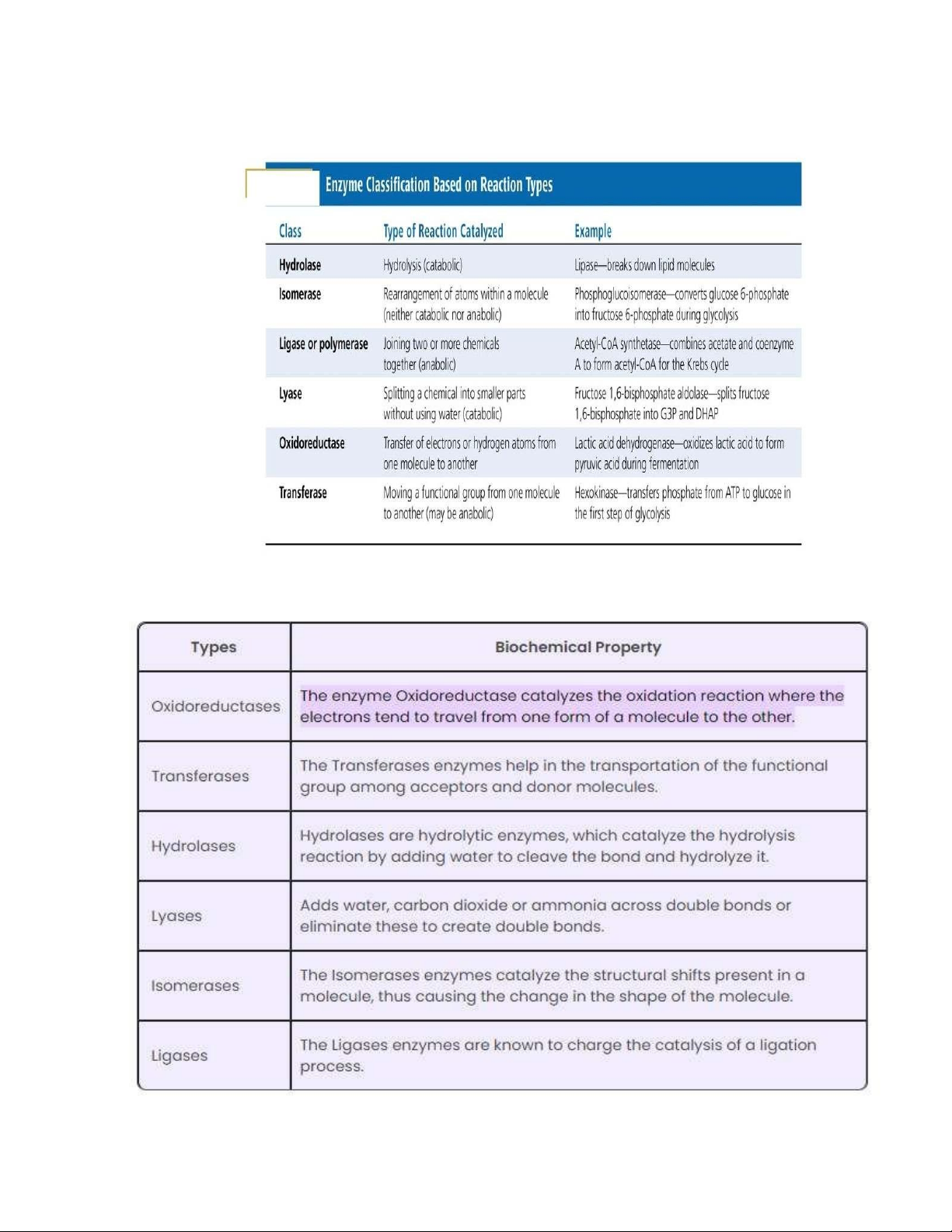
Có bao nhiêu loại Enzyme và Chức năng của chúng lOMoAR cPSD| 59078336
Tài liệu liên quan:
-
Final Exam Notes - Genetic Variability & Development | Môn Biology - Trường Đại học Quốc tế, Đại học Quốc gia Thành phố Hồ Chí Minh
127 64 -
Lab Report 4: Enzyme Activity of Amylase and Catalase in Biology | Môn Biology - Trường Đại học Quốc tế, Đại học Quốc gia Thành phố Hồ Chí Minh
106 53 -
Lecture 5: Understanding Genetics I - Cell Cycle & Mitosis | Môn Biology - Trường Đại học Quốc tế, Đại học Quốc gia Thành phố Hồ Chí Minh
117 59 -
Biology Lab Manual - January 2024 Edition | Môn Biology - Trường Đại học Quốc tế, Đại học Quốc gia Thành phố Hồ Chí Minh
132 66